
|
|
100 consecutive cases of papillary cancer - case 061 |
|


Clinical presentation: A 33-year-old woman was referred for evaluation of a nodular goiter. The patient noticed a lump in her neck for 18 months.
Palpation: a hard, not freely moveable nodule in the right lobe.
Hormonal examination: subclinical hypothyroidism with TSH-level 6.73 mIU/L, FT4 15.6 pM/L, aTPO 2 U/mL.
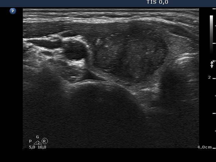
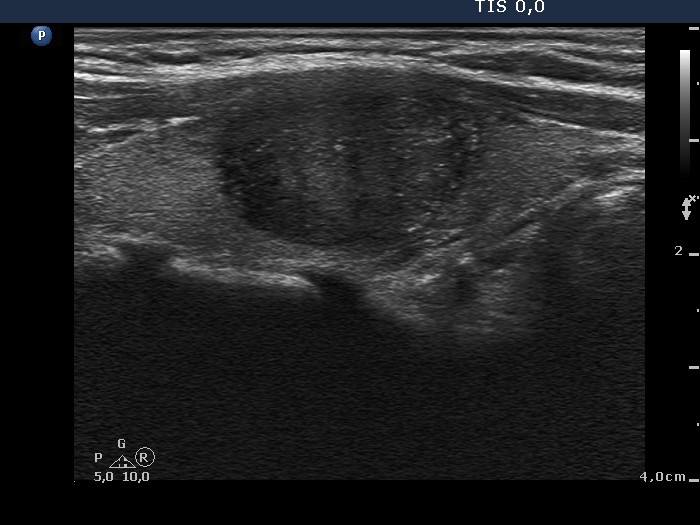
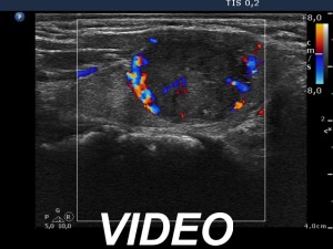
Ultrasonography revealed a hypoechogenic nodule in the right lobe. The lesion showed numerous microcalcifications, had an irregular border and presented perinodular and increased intranodular blood flow.
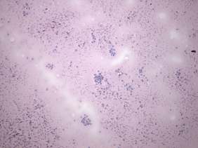
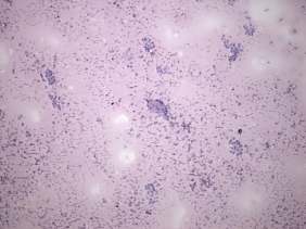
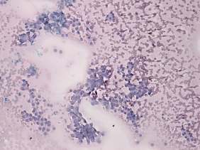
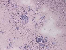
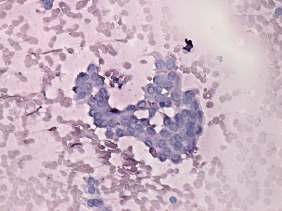
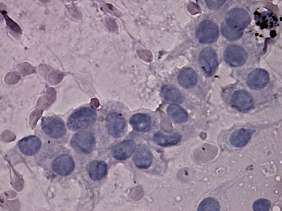
Cytology was performed. There was diffuse colloid in the background. Follicular cells occurred in small, monolayered sheets, in micro- and normofollicles and dissociated. Several enlarged cells were found. Several nuclei contained grooves others had chromatin condensed at the periphery.
Combined cytological-ultrasound diagnosis was suspicion of papillary carcinoma.
Histopathology disclosed papillary carcinoma and chronic lymphocytic thyroiditis.