
|
|
100 consecutive cases of papillary cancer - case 067
|
|


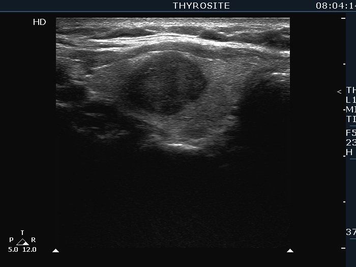


Clinical presentation: A 55-year-old woman was referred for a follow-up examination. We first examined the patient 18 years ago when we saw a discrete lesion with the dimensions of 7x7x9 mm, width x depth x length, respectively.
Palpation: no abnormality.
Functional state: euthyroidism (TSH 0.45 mIU/L).
Ultrasonography. The thyroid was echonormal. There was a hypoechoic nodule in the right lobe. The lesions presented undulation on its surface, but based on the degree of unevenness, the margins cannot be clearly defined as irregular. The dimensions of the nodule were 15x13x17 mm, width x depth x length, respectively. The nodule grew from 0.18 mL to 1.73 mL, i.e. its volume increased almost tenfold.
Cytology resulted in suspicion of papillary carcinoma.
Histopathology disclosed papillary carcinoma.
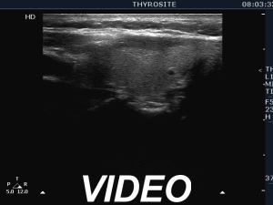
Comment. It is worth comparing the still images with the video. The margins seem to be blurred on still image, but there is a technical reason for this, the image was not stopped at the right moment. The video proves that the borders of the nodule are not blurred.