
100 consecutive cases of papillary cancer - case 068
Follow-up investigation seven years later (ultrasonographic picture 3b)
|
|

|
|
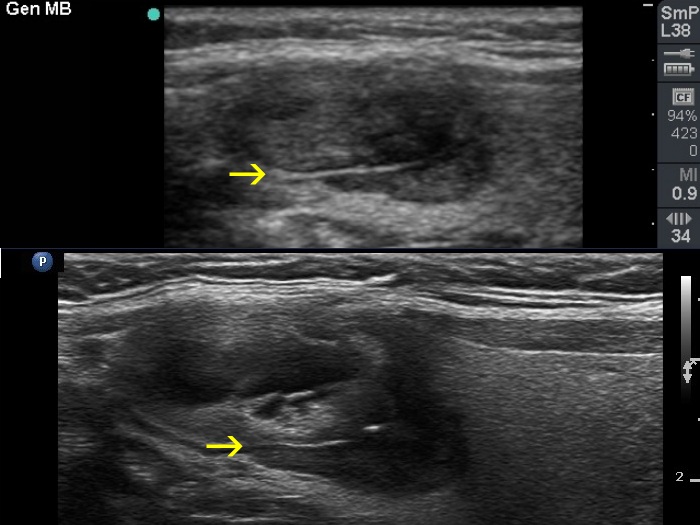
Right lobe, longitudinal scan. Comparison of the ultrasound pattern at first (upper image) and follow-up examination (lower image). Note that the nodule seen as the follow-up clearly as a distinct nodule had been already present at the first examination. However, at this time it did not differ in echogenicity from the larger nodule and was much smaller. Yellow arrows point to a connective tissue fiber which is present at both occasions.