
100 consecutive cases of papillary cancer - case 074 (ultrasonographic picture 2)
|
|

|
|
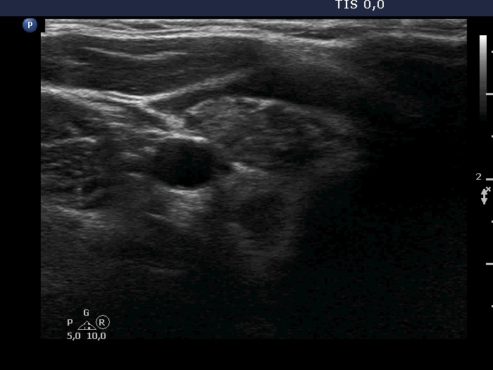
Lower pole of the right lobe, transverse scan. The more hypoechoic area is the lesion in question. In this image it is difficult to judge.