
100 consecutive cases of papillary cancer - case 076
Follow-up investigation a year after the surgery (ultrasonographic picture 3)
|
|

|
|
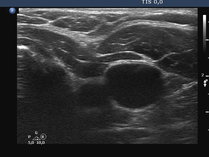
Upper part of the left lobe, transverse scan. There is an echonormal area in the ventral part of the bed. The dorsal anechoic mass is a vessel.