
100 consecutive cases of papillary cancer - case 081 (ultrasonographic picture 3)
|
|

|
|
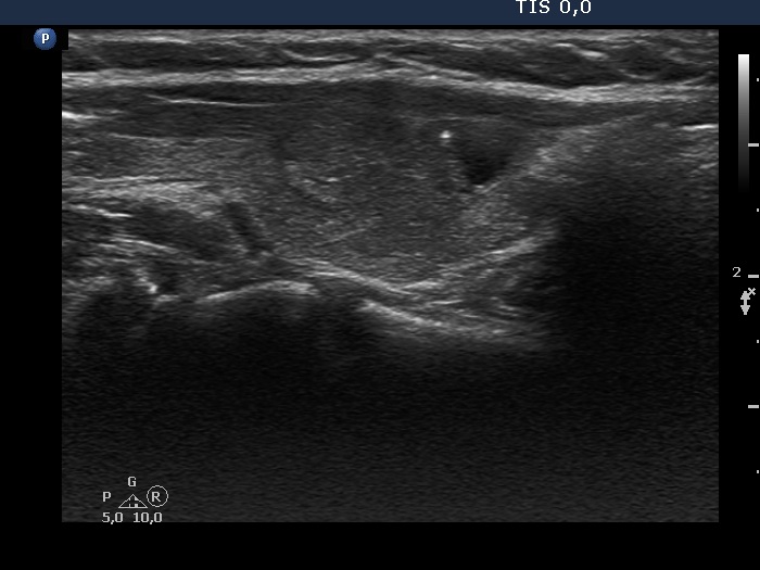
Right lobe, another longitudinal scan. The nodule has lobulated margins. The bright echogenic figure is either a comet-tail artifact of a microcalcification.