
|
|
100 consecutive cases of papillary cancer - case 086
|
|


Clinical presentation: A 58-year-old man was evaluated on fatigue and progressive weight loss. CT scan revealed a vague mass in the lower lung lobe, thereafter PET-CT scan was performed and an incidental focal FDG uptake in the right thyroid was the only positive finding.
Palpation: There was a hard nodule in the right thyroid.
Laboratory test: TSH 1.07 mIU/L.
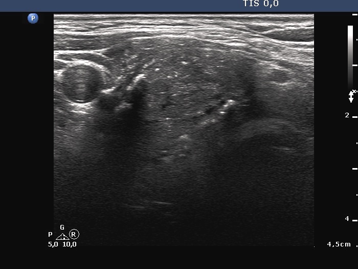
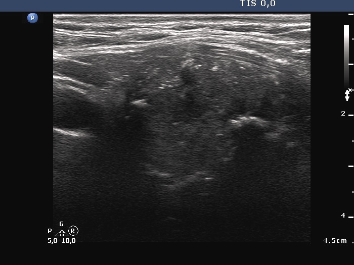
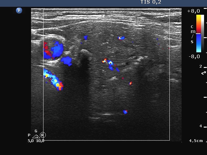
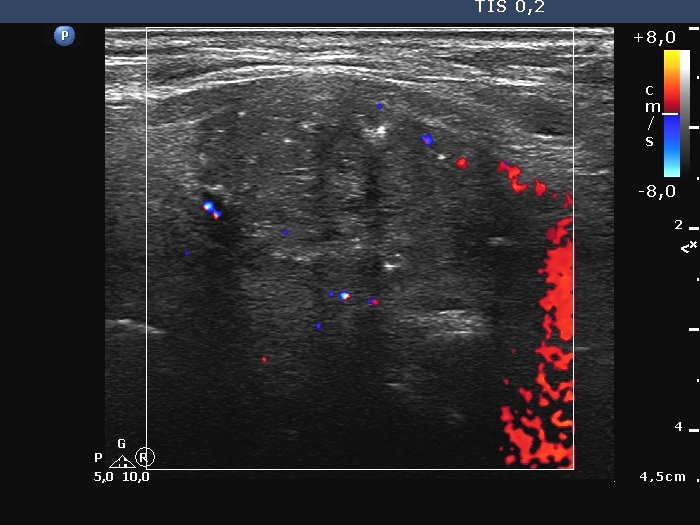
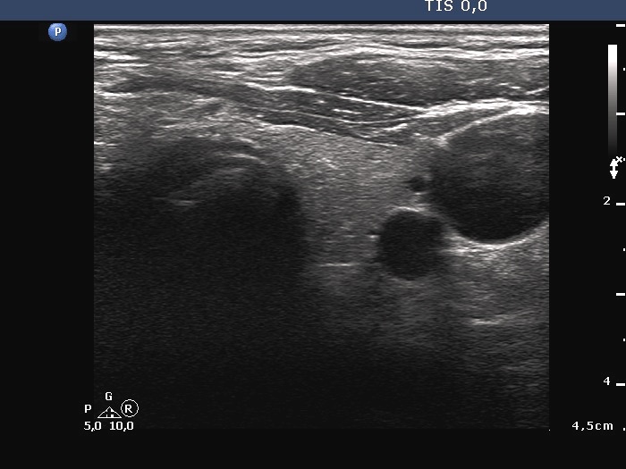
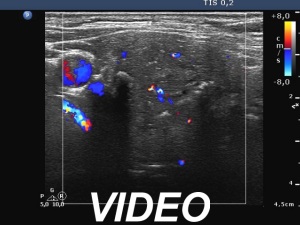
Ultrasonography. The thyroid was echonormal. A large hypoechoic nodule occupied almost the entire right lobe. The nodule showed various intranodular echogenic figures, including microcalcifications, macrocalcification and non-specific figures. The borders were indistinctive. The lesion was suspicious spreading extrathyroidal.
Cytology resulted in papillary cancer.
Histopathology disclosed papillary carcinoma.