
|
|
100 consecutive cases of papillary cancer - case 092
|
|


Clinical presentation: A 57-year-old woman was referred for evaluation of a thyroid nodule which was discovered by the GP.
Palpation: a hard nodule in the right lobe.
Laboratory tests: TSH 3.51 mIU/L, FT4 13.8 pM/L, aTPO < 10 U/mL.
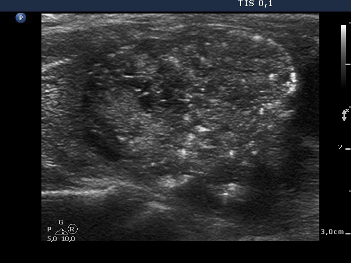
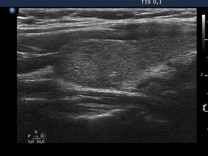
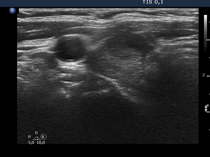
Ultrasonography. The thyroid was echonormal. There were two nodules in the right lobe. The larger palpable one moderately hypoechoic had microcalcifications and irregular borders. There was a smaller, dominantly cystic nodule lower to the former.
On the presence of irregular clusters of follicular cells and small number of grooves cytology alone corresponded only to a Bethesda 3 (atypia of undetermined significance) category. Neither papillary formations nor inclusions wre present.
A combined sonographic-cytological diagnosis of suspicion of papillary cancer was given and total thyroidectomy was suggested.
Total thyroidectomy and right neck lymphadenectomy were performed. Histopathology disclosed papillary cancer.