|
|||||||||||||||||||||||||||
|
Natural course of the disease. Table 8 - Case 26
|
|||||||||||||||||||||||||||
|
|||||||||||||||||||||||||||
|
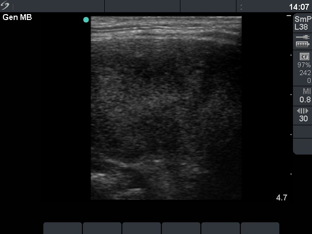
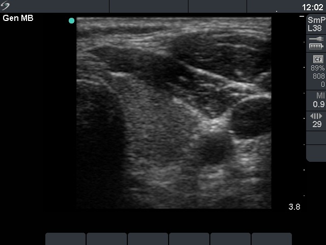
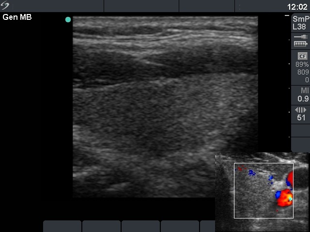
Compare the two series of pictures. There are two main differencies. Firstly, the hypoechogenicity is almost completely resolved at the time of the follow-up examination, and secondly, the size of the thyroid had normalized by that time. Note that the depth was set at 4.7 cm at the first, while at 3.8 cm at the second examination except for the first image. It means that the difference in volume is the best way to compare the first pictures. |
|||||||||||||||||||||||||||