|
|||||||||||||||||||||
|
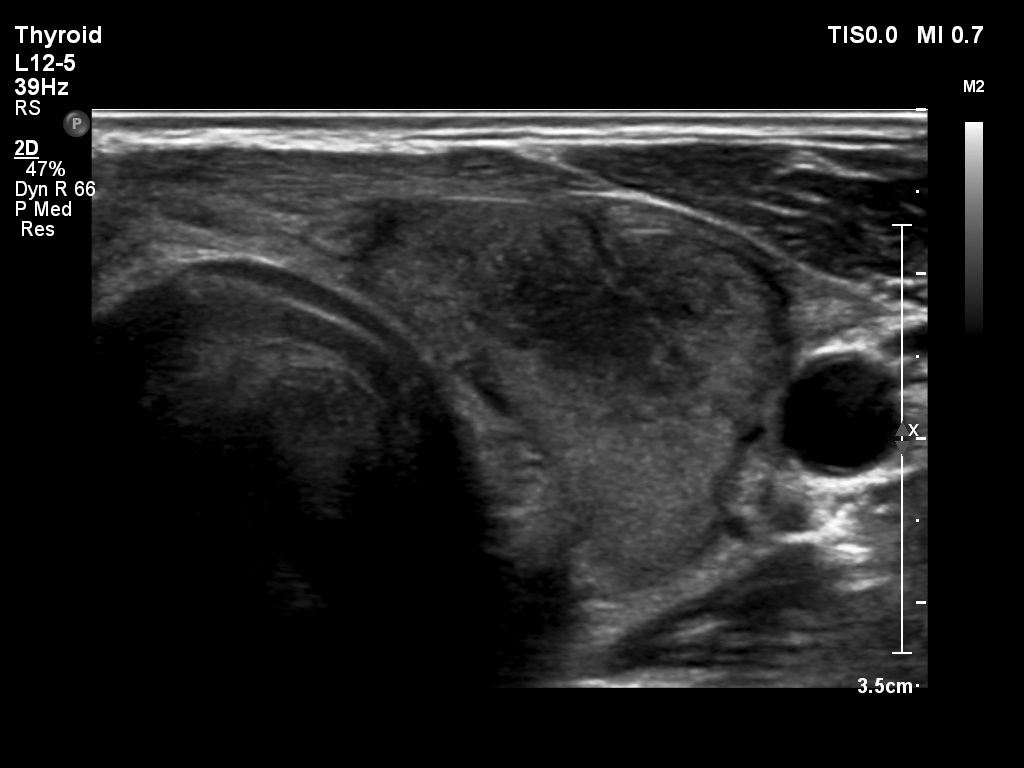
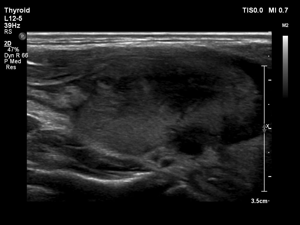
Typical ultrasonographic appearance of the disease. Table 1 - case 43
|
|||||||||||||||||||||
|
|||||||||||||||||||||
|
The most important keys to the diagnosis of de Quervain's thyroiditis are the patchy hypoechogenic areas with ill-defined borders within an echonormal background and the decreased vascularity. Moreover, the inflammated parts of the thyroid are hard on elastography which is demonstrated in the lowest row. The right image is very edifying, here we can see echonormal and hypoechogenic areas. The thyroid is relatively soft corresponding to the former while hard corresponding to the latter. |
|||||||||||||||||||||