Lymph nodes in the neck - Figure 1. How to idetify a lymph node?
The differential diagnostics of a hypoechogenic mass in the neck includes lymph node, a thickened muscle fiber and vessels. If we investigate and we have to investigate a patient in two perpendicular sections then a muscle fiber and a vessel is presented as a pipe-like figure in one scan while a lymph node as a circumscribed oval lesion in all scans. Naturally, Dopple mode is decisive itself in the case of a vessel.
Differentiation of a lymph node from a muscle fiber. Lymph nodes in the neck - case 12 |
|
 |
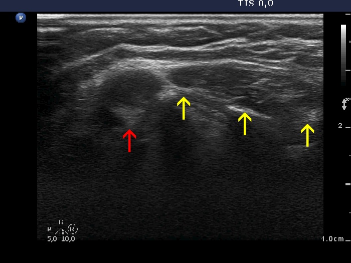 |
There are two hypoechogenic masses in the left, horizontal view. The perpendicular, longitudinal section (right image) is decisive. The lymph node marked with a red arrow is a relatively small circumscribed lesion with oval shape while the muscle fiber marked with yeloow arrows runs along the transducer and has a pipe-like shape. |
|
Differentiation of a lymph node from vessels. Lymph nodes in the neck - case 7 |
|
 |
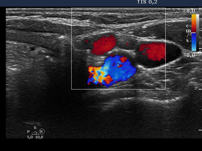 |
There is a lymph node among three vessels. The node presents a less hypoechogenic structure. If we had any doubt, the Doppler mode is decisive.