|
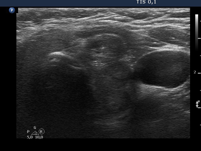
Transverse scans |
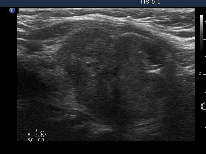
Longitudinal scans |
 |
 |
 |
 |
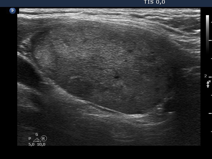
This is a heterogeneous, dominantly echonormal nodule. The echogenicity of the nodule is significantly lower on longitudinal scan than on transverse view. This is caused by technical problem: the transducer could not be fit to appropriately to the neck of the patient which led to decrease in echogenicity.
|
|
|
Hashimoto's thyroiditis (cytology) - case 2168 |
 |
 |
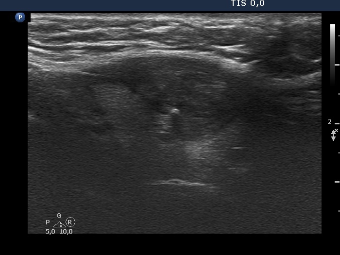
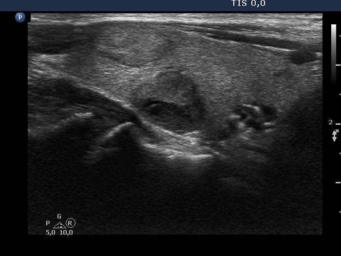
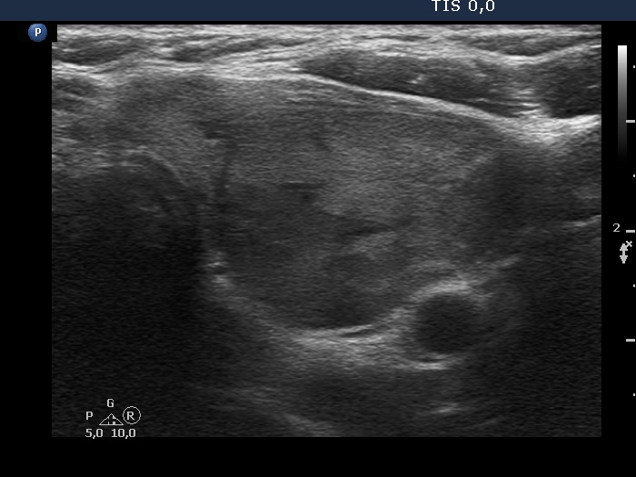
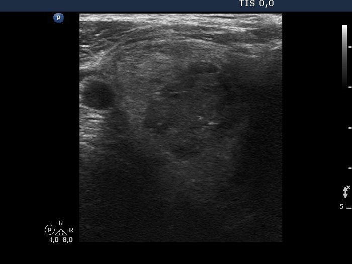
Although there are tiny hypoechoic portions within the nodule, the proportion of them is below 10% and the largest diameter of the hypoechoic part is less than 1 cm. Therefore, this nodule should be called homogeneous. Moreover, the hypoechoic part within the nodule is very likely the presentation of the infiltration of the underlying thyroiditis.
|
|
|
Benign hyperplastic nodule (histology) - case 2050
|
 |
 |
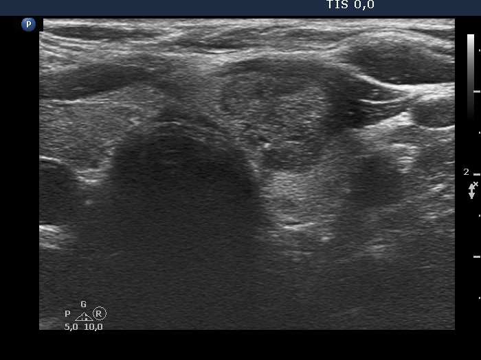
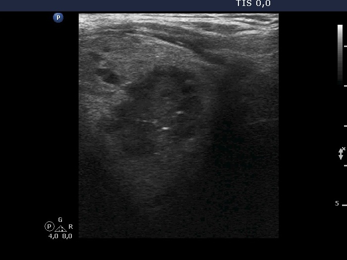
The nodule with irregular borders is composed of dominantly moderately hypoechoic portions and has less hypoechoic parts. If the latter are counted as minimally hypoechoic than the nodule does not belong to heterogeneous lesions, because both the minority and the dominant portions belong to the same subgroup of echogenicities, to the hypoechoic one. If we judge the less hypoechoic parts as echonormal, that the nodule should be counted as heterogeneous.
|
|
Metastasis of a laryngeal carcinoma to the thyroid - case 2110
|
Transverse scan |
Longitudinal scan |
 |
 |
 |
 |
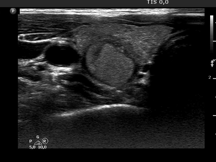
The nodule is dominantly hypoechoic and has small portions which are minimally hypoechoic. Note that these areas (yellow arrow) are darker than the extranodular part of the thyroid (red arrow). It means that the nodule is not heterogeneous (echonormal-hypoechoic) but is hypoechoic. It is not evident whether the echogenicity of the hypoechoic part is lighter or darker compared to the strap muscle fibers.
|
|
|
|
Transverse scan |
Longitudinal scan |
 |
 |
 |
 |
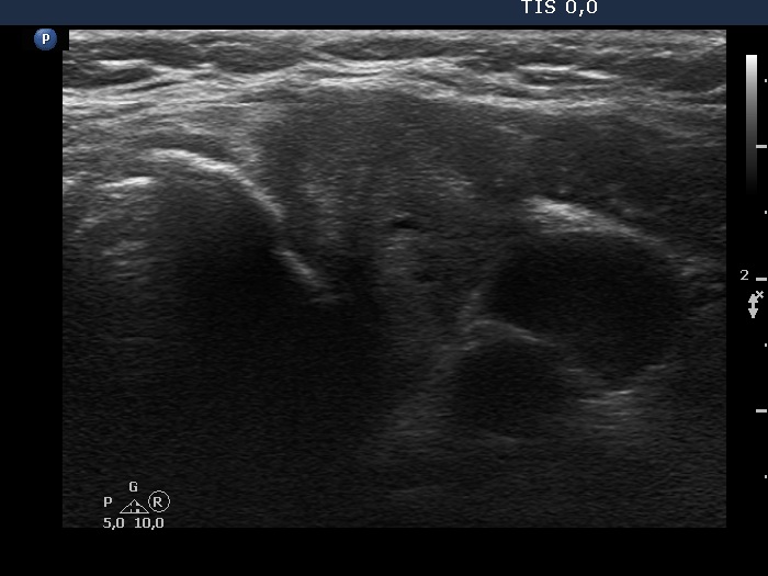
The dominant part of the dorsal nodule is minimally hypoechoic while the minority part is less dark. The dorsal nodule is either homogeneously hypoechoic (i.e. composed of deeply and minimally hypoechoic parts) or heterogeneous (i.e. composed of hypoechoic and echonormal parts) depending on the judgement of the less dark portion of the nodule.
|
|
|
|
 |
 |
This is a dominantly echonormal, heterogeneous nodule.
|
|
|
|
|
 |
 |
 |
 |
These are dominantly hypoechoic, heterogeneous nodules. The dominant part is moderately hypoechoic in the left while deeply hypoechoic in the right case.
|
|
|
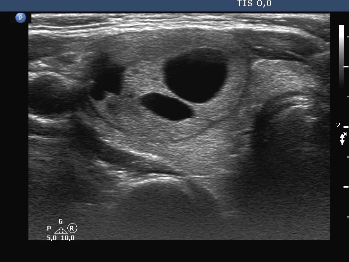
Benign cystic-colloid goiter (cytology) - case 2111 |
 |
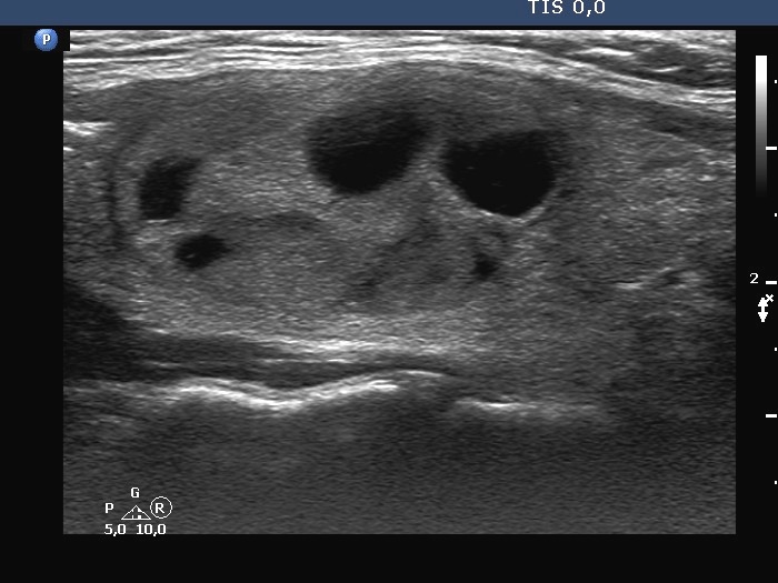 |
This is a mixed, dominantly solid nodule. The solid part is heterogeneous. The solid part contains almost equal proportion of echonormal and minimally/moderately hypoechoic parts.
|
|
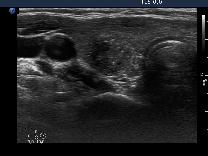
Benign cystic-colloid goiter (cytology) - case 2176 |
 |
 |
This is a mixed, dominantly cystic nodule. Although the solid part has parts with different echogenicities, it can be ecxluded that thi minority hypoechoic portion can have different pathology than the dominant, isoechoic part. SO thie solid part is homogeneously echonormal.
|
|
|
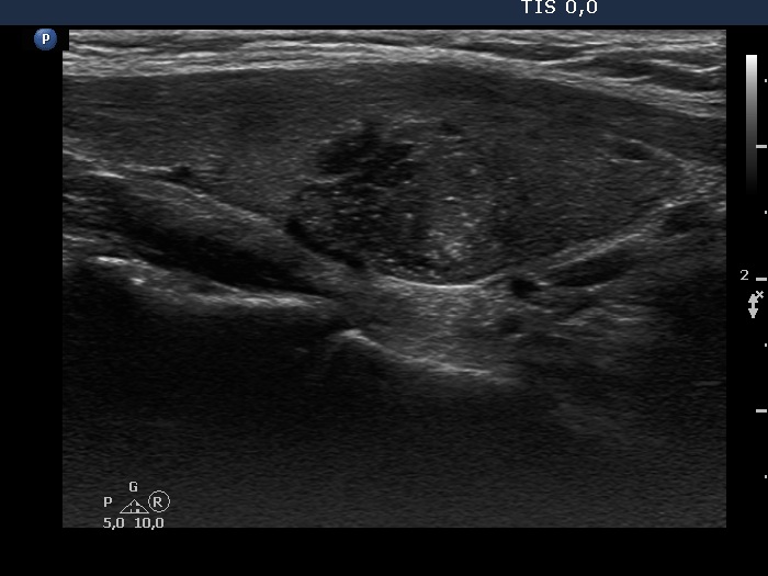
Papillary carcinoma in Hashimoto's thyroiditis (histology) - case conp 033 |
 |
 |
The dominantly echonormal nodule has hypoechoic areas. Nevertheless, these occur at the border of the lesion and very likely represent the infiltration of the nodule by the underlying thyroiditis. Therefore, the nodule is not a heterogeneous lesion.
|
|
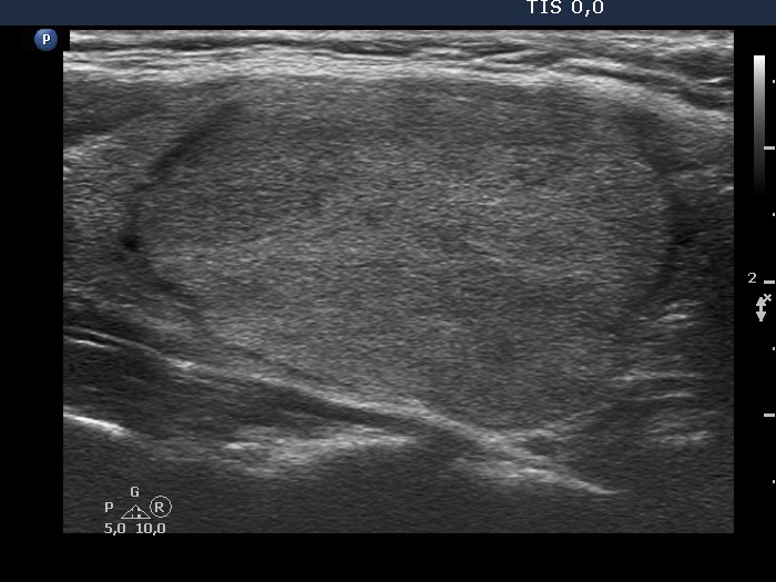
Papillary carcinoma in Hashimoto's thyroiditis (histology) - case conp 054 |
Transverse scan |
Longitudinal scan |
 |
 |
The lesion in the ventral part of the right lobe shows a very unusual presentation: it is composed of a central hypoechogenic area surrounded with an echonormal rim.
|
|
Benign nodule in Hashimoto's thyroiditis (cytology) - case 782 |
Transverse scan |
Longitudinal scan |
 |
 |
The lesion in the right lobe has a heterogeneous pattern. The peripheral parts of the nodule are hypoechoic which raises the possibility that the nodule is not a heterogeneous lesion, but an echonormal nodule which is infiltrated by the underlying thyroiditis.
|
|
|
Benign nodule in Hashimoto's thyroiditis (cytology) - case 1401 |
Transverse scan |
Longitudinal scan |
 |
 |
The lesion in the right lobe has a heterogeneous pattern. The peripheral parts of the nodule are hypoechoic which raises the possibility that the nodule is not a heterogeneous lesion, but an echonormal nodule which is infiltrated by the underlying thyroiditis.
|
|
|
Papillary carcinoma in Hashimoto's thyroiditis (cytology) - case 218 |
Transverse scan |
Longitudinal scan |
 |
 |
The tumor has a heterogeneous pattern. The upper-lateral part of the nodule is hypoechoic while the dominant part of the lesion is isoechoic, i.e. it has a similar pattern to the extranodular portion of the lobe. However, if we compare the nodule' echogenicity to a healthy thyroid than all parts of the lesion are hypoechoic.
|
|
Discrete lesion in Hashimoto's thyroiditis (cytology) - case 2067 |
Transverse scan |
Longitudinal scan |
 |
 |
The lesion in the right lobe has a heterogeneous pattern. It seems more likely that this is not a real nodule, but a relatively intact part of the lobe that has not yet been completely infiltrated by inflammation.
|
|
Follicular carcinoma (histology) - case 68 |
Transverse scan |
Longitudinal scan |
 |
 |
This is a dominantly isoechoic nodule which presents minimally hypoechoic islets.
|