 |
|
 |
Table 2 Histogram analysis
|
Histogram analysis is a quite simple method which can be the basis for the exact determination of the nodule' echogenicity. We present here three examples which demonstrate the importance of the definitions. Unfortunately, the definition of the reference points is either not clear (muscle tissue) or not uniform (normal thyroid) in the literature which might explain the low interobserver agreement on the echogenicity.
Oxyphilic adenoma (histology) - case 60 |
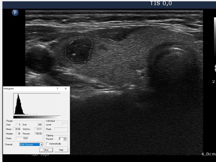
Histogram analysis of the nodule
|
 |
 |
Mean 29.0 |
Mean 28.0 |
This homogeneous nodule three echogenic granules which accounted less than 2% of the surface of the nodule, there for the grey-level of the entire nodule and that of those parts which did not involve the echogenic granules were almost the same.
|
Histogram analysis of the strap muscle
|
 |
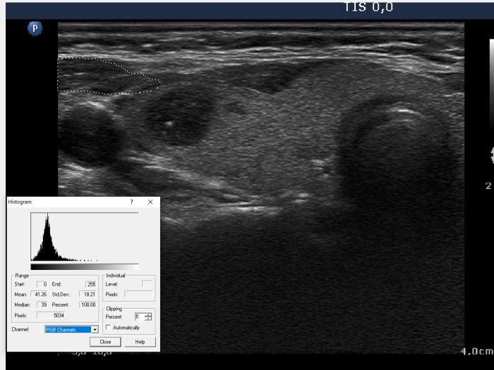 |
Mean 31.0 |
Mean 41.3 |
The gray-level of the entire muscle exceeds that of the muscle fiber by more than 30%
|
Histogram analysis of the normal thyroid
|
The entire nod-nodular part |
The ventral third of a specific section |
 |
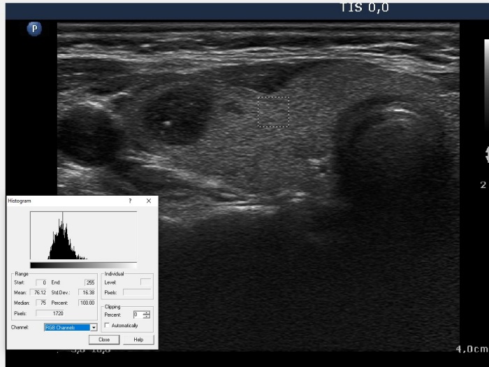 |
Mean 66.3 |
Mean 76.1 |
Histogram analysis of the normal thyroid
|
The middle third of specific section |
The dorsal third of a specific section |
 |
 |
Mean 73.2 |
Mean 64.2 |
Note that the grey level of the normal parenchyma decreases with depth. The most ventral third exceeds the grey level of the most dorsal third by around 20%.
|
|
|
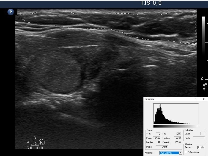
Benign nodule in Hashimoto's thyroiditis (cytology) - case 851 |
Transverse scan |
Longitudinal scan |
 |
 |
|
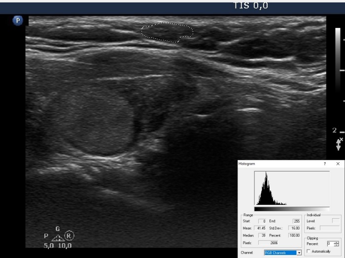 |
The histogram analysis of the nodule resulted in 48.6. The grey-level of the entire muscle was 51.3, while that of the muscle fiber was 41.5. It means that depending on the definition of the strap muscle, the nodule is either deeply hypoechoic or not.
|
|
 |
|
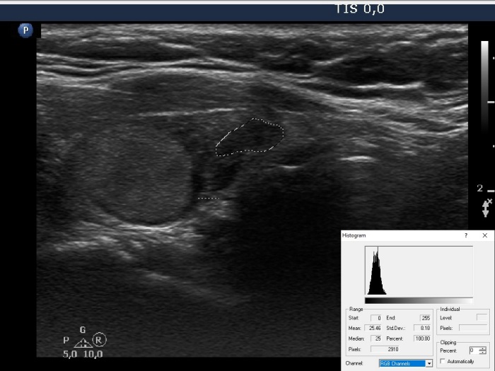 |
The histogram analysis of the nodule resulted in 48.6. The average grey-level of the non-nodular thyroid was 42.0, while that of the dominant part of the non-nodular part was 25.5. I summarized in the table below the classification of the nodule depending on the reference tissues taken into account.
|
Depending on the reference tissues, the nodule can be considered either as hyperechoic or moderately hypoechoic or deeply hypoechoic.
Definition of the reference tissues |
Judgement of the echogenicity of the nodule (PEV=48.6) |
The normal thyroid |
The strap muscle |
The entire extranodular part (PEV=42.0) |
The entire strap muscle (PEV=51.3) |
Hyperechoic and deeply hypoechoic at the same time |
The portion of the extranodular part with the dominant echo structure (PEV=25.5) |
Hyperechoic and deeply hypoechoic at the same time |
The mean of 100 healthy thyroids (PEV=71.9) |
Deeply hypoechoic |
The entire extranodular part (PEV=42.0) |
The muscle fiber (PEV= 41.5) |
Hyperechoic |
The portion of the extranodular part with the dominant echo structure (PEV=25.5) |
Hyperechoic |
The mean of 100 healthy thyroids (PEV=71.9) |
Moderately hypoechoic |
PEV means the mean pixel echogenicity value which ranges theoretically from 0 to 255, from the darkest to the brightest level, respectively.
|
|
|
|
Histogram analysis of a benign nodule in Hashimoto's thyroiditis (cytology) - case 1782 |
 |
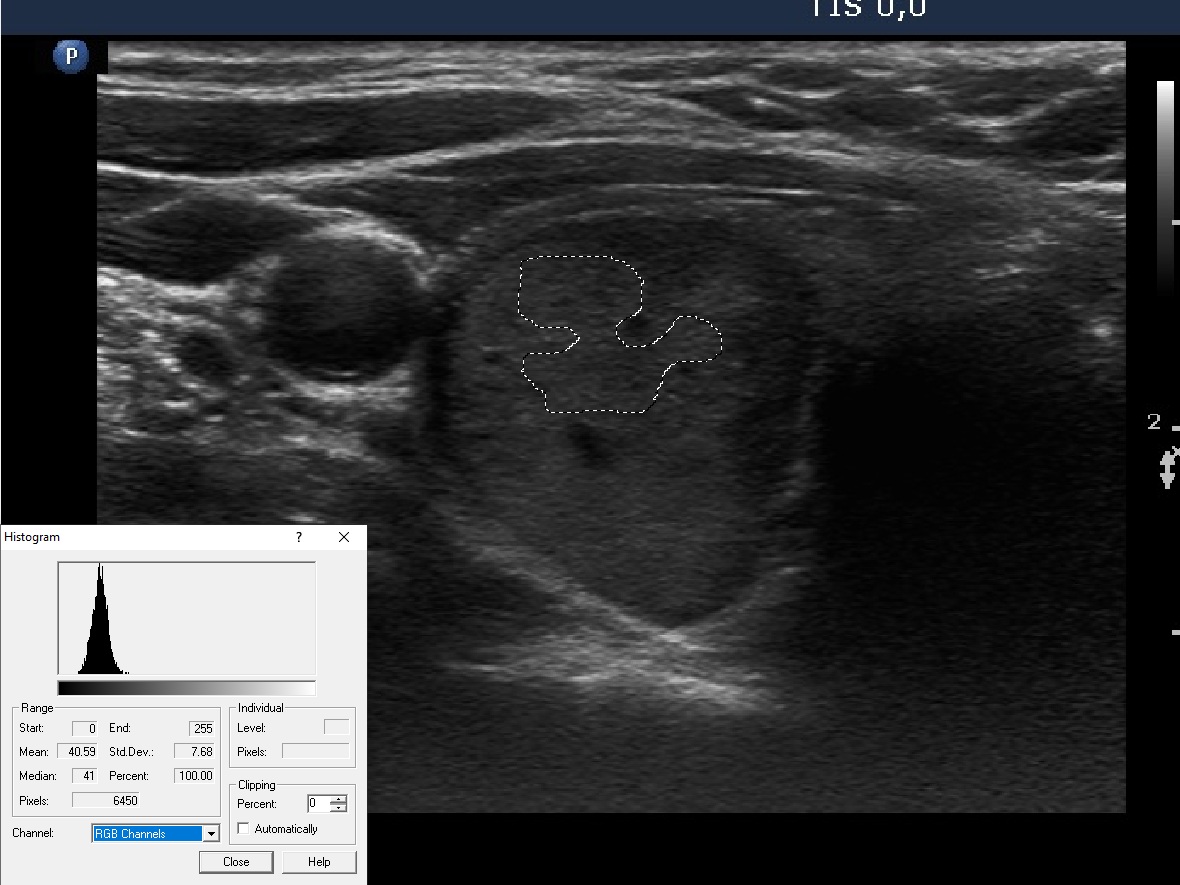 |
At first glance, the nodule seemed more echogenic than the extranodular part to me. Thereafter, I have measured the histogram values, and the nodule proved to be less echogenic: the histogram value of the dominant part of the nodule is 40.6, the histogram value of the extranodular part if 44.8. It means that the nodule is hypoechoic.
|
| |
|
|