| |
|
Measuring of the histogram values |
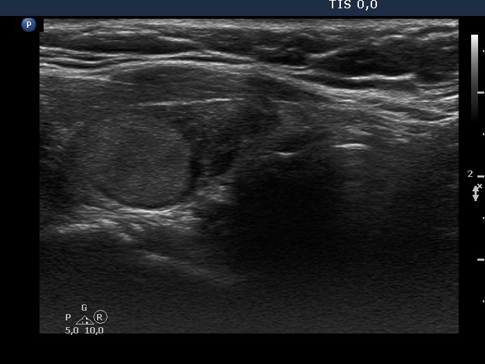
The normal image of the right lobe with a nodule

|
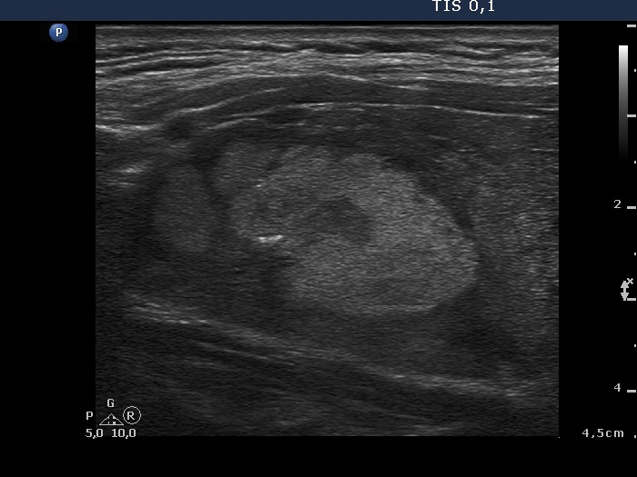
The histogram of the nodule (while circumscribing the portion with the dominant echogenicity, hyperechogenic figures and if present cystic areas are excluded)

|
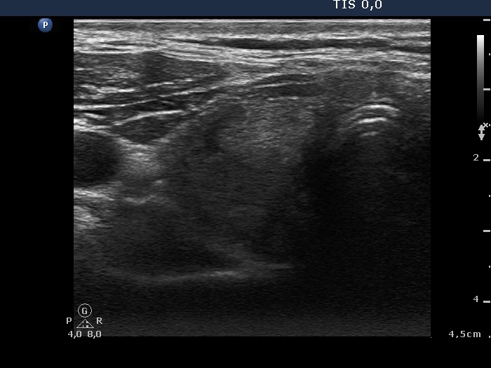
The histogram of the brightest part of the non-nodular parenchyma

|
The histogram of the muscle fibers of the strap muscle

|
The histogram value of the nodule was 45.7, which was between the value of the normal parenchyma 86.5) and that of the muscle fiber (37.5). It means on the one hand, that the nodule was darker than the non-nodular part, i.e. the nodule proved to be hypoechoic. On the other hand, the nodule was brighter than the muscle fiber, i.e. the lesion did not prove deeply hypoechoic. As a result, the nodule was minimally/moderately hypoechoic.
|
|
|
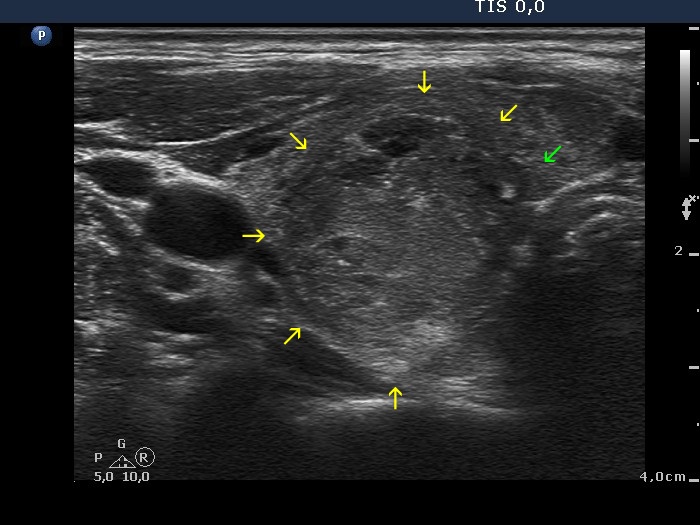
Oxyphilic adenoma in Hashimoto's thyroiditis (histology) - case 554 |
|
|
 |
 |
 |
 |
Although the non-nodular part is also hypoechoic, the dominant part of the nodule is more hypoechoic. It means that irrespectively of the reference tissue, the dominant part of the nodule should be considered hypoechoic. The reference tissue has an influence whether this lesion should be considered as a dominantly hypoechoic, heterogeneous nodule or simply as a hypoechoic one because the echogenicity of the brighter islets within the nodule (right image, histogram value 64.9) are more echoic than the extranodular part (left image, histogram value 46.9) but less echoic than a healthy thyroid.
|
|
|
Hashimoto's thyroiditis (cytology) - case 1749 |
Transverse scans |
Longitudinal scans |
 |
 |
The thyroid is minimally-moderately hypoechoic. There is a mixed nodule in the upper third of the lobe. The issue is the interpretation of the solid part. It is clearly more echogenic than the non-nodular part. If we compare the nodule' echogenicity to the non-nodular part than this lesion is hyperechoic. However, the histogram value of the solid part (54.9) is between that of the average of the normal thyroid tissue (82.0) and muscle fiber (36.6). It would mean that the solid part is minimally/moderately hypoechoic.
|
|
Benign nodule is Hashimoto's thyroiditis (cytology) - case 851 |
Right lobe |
Left lobe |
 |
 |
 |
 |
The histogram value of the nodule in the right lobe is 64.6, while this value proved to be 49.0 in the event of the nodule in the left lobe. So, if we define nodule' echogenicity in relative sense than both nodules are iso/hyperechogenic, if we define nodule echogenicity in absolute sense than both nodules are minimally/moderately hypoechoic.
|
|
|
Hashimoto's thyroiditis without any nodules (histology) - case 477
|
 |
 |
The histogram value of the lesion in the right lobe is 105.6. So, irrespectively of the reference tissue used, this lesion is hyperechoic.
|
|
Benign lesion in Hashimoto's thyroiditis (cytology) - case 24
|
Transverse scan |
Longitudinal scan |
 |
 |
If we compare the nodule to the extranodular part than the lesion is hyperechoic. However, if the reference tissue is a healthy thyroid than the nodule should be classified as moderately hypoechoic.
|
|
|
|
Transverse scan |
Longitudinal scan |
 |
 |
The histogram value of the lesion is 69.4. It means that although the nodule is more echogenic than the-non-lesional part of the lobe, its echogenicity is lower compared with a healthy thyroid. The latter is characterized by a mean histogram value of 82.0. So, the nodule should be grouped among iso/hyperechogenic lesions if the reference tissue is the non-nodular part while should be grouped among moderately hypoechogenic nodules if the reference tissue is the healthy thyroid.
|
|
|
Hashimoto's thyroiditis (cytology) - case 1620 |
 |
 |
The histogram value of the extranodular part of the right lobe is 23.6 while that of the discrete lesion is 31.6. It means that if the reference tissue is the non-nodular part than the lesion is hyperechoic. On the other hand, if we compare the echogenicity of the lesion to a healthy thyroid than this is a moderately hypoechoic lesion.
|
|
|
Hashimoto's thyroiditis (cytology) - case 1019
|
 |
 |
The histogram value of the discrete lesion is 52.0 while that of the extranodular part is 45.8. The categorization of the nodule depends on what to compare the echogenicity to. If the reference is the non-lesional part of the thyroid than the lesion is hyperechoic. However, compared to a healthy thyroid, the nodule is hypoechoic.
|
|
|
Hashimoto's thyroiditis (cytology) - case 793 |
 |
 |
The histogram value of the extranodular part of the right lobe is 36.1 while that of the discrete lesion is 64.0. It means that if the reference tissue is the non-nodular part than the lesion is hyperechoic. On the other hand, if we compare the echogenicity of the lesion to a healthy thyroid (which is characterized by a 82.0 histogram value) than this is a moderately hypoechoic lesion.
|
|
Papillary carcinoma in Hashimoto's thyroiditis (histology) - case conp033 |
 |
 |
The histogram value of the nodule proved to be 74.8, a value which is less than the average of the normal parenchyma, therefore in absolute sense this is a minimally hypoechoic nodule. In relative sense, compared to the non-nodular part of the particular case, the nodule is hyperechoic.
|
|
|
Hyperplastic nodules in Hashimoto's thyroiditis (histology) - case 54 |
Transverse scans |
Longitudinal scans |
 |
 |
 |
 |
These discrete lesions are not heterogeneous ones. The hypoechoic areas within are the presentations of the infiltration of the underlying thyroiditis.
|
|
Hashimoto's thyroiditis (cytology) - case 479 |
 |
 |
The situation is similar to the previous case. The discrete lesion is infiltrated by the underlying thyroiditis. The histogram value is 84.7. It means that the lesion belongs to the iso/hyperechogenic nodules both in the relative and absolute term, compared to the non-nodular part and to the healthy thyroid, respectively.
|
|
|
Hashimoto's thyroiditis (cytology) - case 104 |
 |
 |
The entire lesion is brighter than the extranodular part. However, the less bright parts of the nodule are hypoechoic if we compare the echogenicity to a healthy thyroid. It means that if the reference tissue is the extranodular part than the lesion should be classified as hyperechoic while if the reference tissue would be a healthy thyroid than this lesion is a dominantly iso/hyperechoic nodule which has moderately hypoechoic parts.
|
|
|
Hashimoto's thyroiditis (cytology) - case 95 |
 |
 |
The categorization of the isoechoic nodule in the left lobe depends on the definition of normal thyroid. If it is the extranodular part than the nodule is iso/hyperechoic. If the reference tissue is the healthy thyroid than the nodule is minimally/moderately hypoechoic because the histogram value of the nodule (56.0) is lower than that of the healthy thyroid (82.0).
|
|
|
Hashimoto's thyroiditis (cytology) - case 1739 |
 |
 |
This nodule is more hypoechoic than the extranodular tissue. However, the echogenicity is of the lesion is less bright than a normal, healthy thyroid.
|
|
|
Hashimoto's thyroiditis (cytology) - case 61 |
 |
 |
The definition of the nodule depends on what to compare the echogenicity to. If the reference is the non-lesional part of the thyroid than the lesion is hyperechoic. However, compared to a healthy thyroid, the nodule is hypoechoic.
|
|
|
Hashimoto's thyroiditis (cytology) - case 99 |
 |
 |
The histogram value of the nodule is 69.3. The judgement of the echogenicity of the nodule in the left lobe depends on how to define the reference normal thyroid. If it is the extralesional part than the nodule is iso/hyperechoic and according to most TIRADS does not require FNA. Is the reference tissue is the healthy thyroid, than the nodule should be involved among hypoechoic nodules and is a candidate for FNA.
|
|
Papillary carcinoma in Hashimoto's thyroiditis (histology) - case conp 035 |
Transverse scans |
Longitudinal scans |
 |
 |
 |
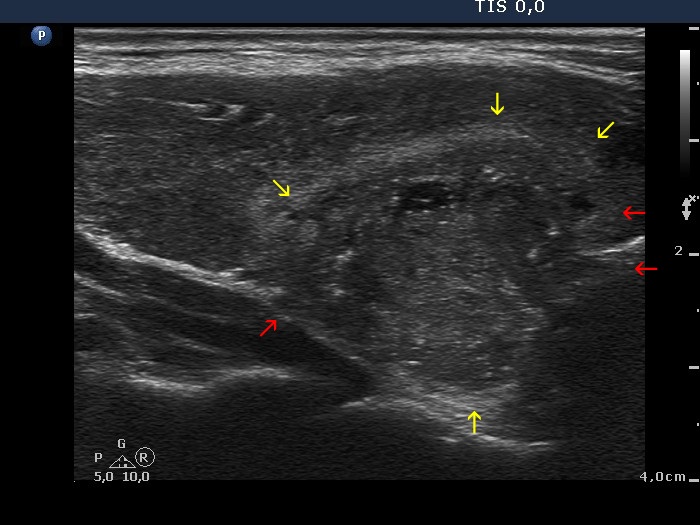 |
The papillary carcinoma (marked with yellow arrows) was located in the dorsal lesion. This differs from the non-tumorous part in the presence of microcalcifications and in the echogenicity. The tumor is less hypoechoic than the surrounding thyroid tissue.
|
|
|
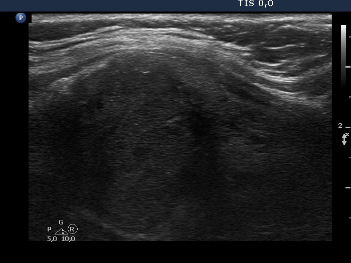
Benign hyperplastic nodule in Graves' disease (histology) - case 1147 |
Examination in hyperthyroid state |
Examination in euthyroid state |
 |
 |
 |
 |
Compare the echogenicity of the nodule (marked with yellow) and that of the extranodular part (marked with red arrows) in hyperthyroid an euthyroid state. In hyperthyroid state the nodule is more echogenic while in euthyroid state the nodule is less echogenic than the extranodular part. In the former state the lesion should be considered hyperechogenic while in the latter the lesion should be considered as hypoechoic.
|
|
|
Hashimoto's thyroiditis (cytology) - case 1749 |
|
|
The judgement of the solid part of the lesion depends on the reference tissue: if it is the extralesional parenchyma than the lesion is echonormal or hyperechoic. If the reference tissue is the healthy thyroid than the lesion is minimally hypoechoic.
|
| |
|
| |
|
| |
|
|