|
 |
 |
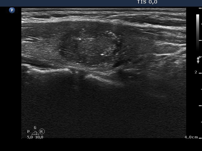
The hypoechogenic areas present both echogenic lines and granules which synchronous presence is the hallmark of connective tissue. Note the multiplicity, the irregular shape and borders of these lesions. All of these properties stand for these lesions being presentations of Hashimoto's thyroiditis. |
| |
|
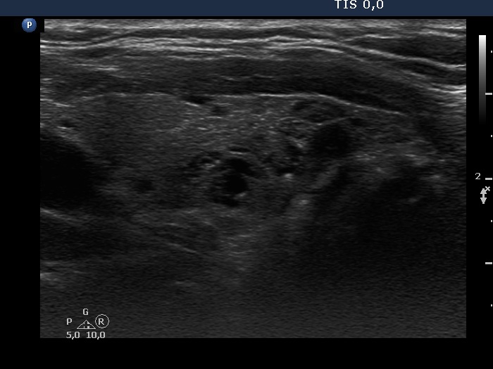
Right lobe, horizontal scan |
Right lobe, longitudinal scan |
 |
 |
Numerous bright granules and lines coexist in the thyroid. These echogenic figures correspond to connective tissue.
|
| |
Benign hyperplastic nodule in a Graves' patient (histology) - case 1673 |
 |
 |
The presence of coarse calcification is a strong argument that the lesion is a true nodule. |
| |
Papillary carcinoma in Graves' disease - case 993 |
Right lobe, horizontal view |
Right lobe, longitudinal view |
 |
 |
There are several bright punctate echogenic foci and less bright granules and lines, as well. The former corresponds to microcalcifications, while the latter does to connective tissue. |
| |
|
Right lobe, horizontal scan |
Right lobe, longitudinal scan |
 |
 |
 |
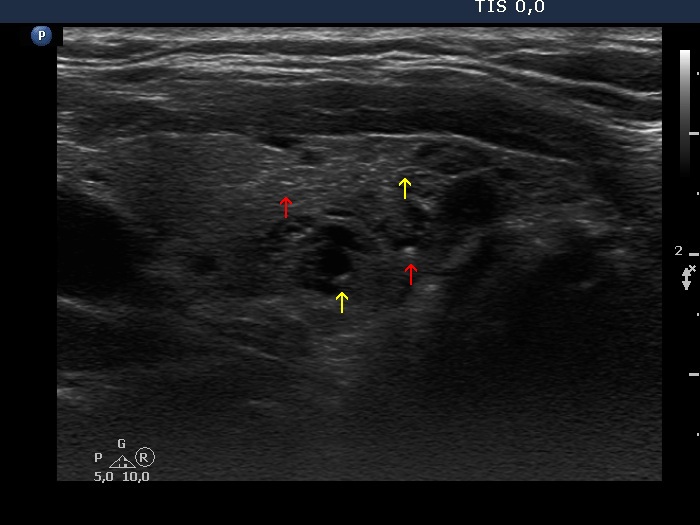 |
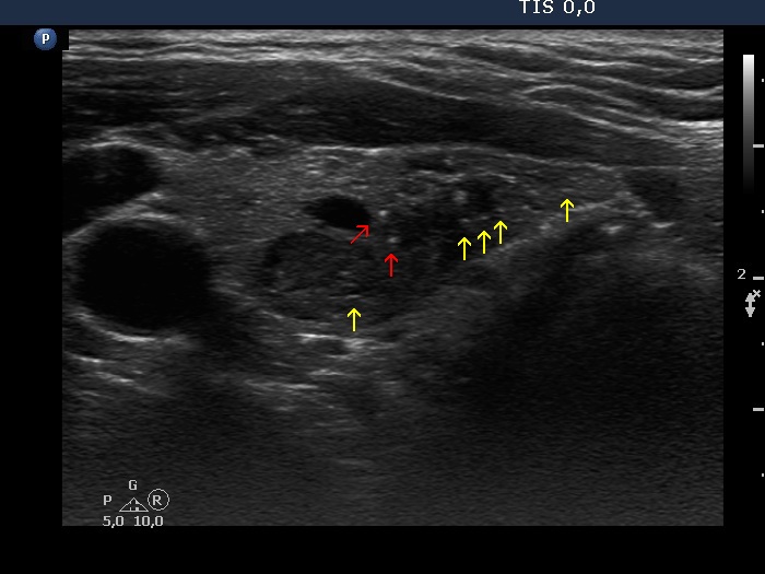
The interpretation of smaller and less bright granules and lines (yellow arrows) is not very difficult, they correspond to connective tissue. On the other hand, there are a few more bright and a bit larger echogenic granules (red arrows). The finding of one similarly bright line in the right image is of help: these figures are also very likely presentations of connective tissue. |
| |
|
Right lobe, horizontal scans |
Right lobe, longitudinal scans |
 |
 |
 |
 |
There is a small hypoechogenic lesion in the lower pole of the right lobe. The lesion presents both echogenic lines and granules; therefore these figures correspond to connective tissue.
|
| |
| |
|
First examination |
Follow-up 18 months later |
 |
 |
 |
 |
The nodule in the dorsal part of the right lobe proved to be papillary carcinoma. It is worth comparing the difference in the ultrasound presentation of the nodule and the extranodular part in the right lower image: the tumor focus presents microcalcifications while the non-nodular part does not. There are no other relevant differences.
|
| |
Graves' disease without any nodules - case 888 |
Papillary carcinoma in Hashimoto's thyroiditis - case conp009 |
 |
 |
 |
 |
The lesion in the lower third of the lobe was described as a suspicious nodule. In fact this is only the largest among the numerous hypoechogenic areas. It has irregular shape and borders which features also stand against a pathological nodule. The lesion has both intralesional granules and lines, the coexistence of which corresponds to connective tissue and not to punctate echogenic foci (microcalcifications).
|
The interpretation of the intranodular echogenic figures is equivocal. Beside echogenic granules we can find echogenic lines as well. However, the latter are a bit less bright.
|
Although the presentations of the above cases differ, first of all because of the infiltrative growth of the malignant focus, cytology was also indicated in the left, benign case. This is not a rare situation in hypoechogenic lesions of Hashimoto's thyroiditis: although a discrete lesion is probably not a nodule, cytology might be indicated on the ultrasound presentation.
|
| |
|
Hashimoto's thyroiditis (histology) - case 441 |
Papillary carcinoma in Hashimoto's thyroiditis - case conp061 |
 |
 |
 |
 |
There is a lesion next upper to a large hypoechogenic nodule (see the lower, longitudinal scan). The small area has echogenic figures which likely belong to echogenic foci (microcalcifications) subgroup. According to this lesion there was no nodule on pathology.
|
The nodule has several microcalcifications.
|
The left case demonstrates the limitations of our knowledge and/or technical modality: a highly suspicious discrete area proved to be only a more active focus of Hashimoto's thyroiditis. Note the striking similarity of the presentations of the two cases.
|
| |
|
Papillary carcinoma in Hashimoto's thyroiditis - case 1589 |
Right lobe, horizontal scan |
Right lobe, longitudinal scan |
 |
 |
The thyroid is composed of discrete hypoechogenic areas. The tumor focus presents punctate echogenic foci, as well. |
| |
|
Papillary carcinoma in Hashimoto's thyroiditis - case conp057 |
Right lobe, horizontal scan |
Right lobe, longitudinal scan |
 |
 |
The small lesion is proved to be papillary carcinoma. The nodule has both macrocalcifications and punctate echogenic foci. The former is a rare finding in a discrete lesion of Hashimoto's thyroiditis.
|
| |
| |
Papillary carcinoma in Hashimoto's thyroiditis - case conp016 |
|
 |
 |
 |
 |
The tumor presents the so-called starry sky phenomenon causes by the numerous microcalcifications.
|
The dorsal, moderately hypoechogenic lesion presents both echogenic lines and granules.
|
| |
Hashimoto's thyroiditis and suspicion of papillary carcinoma (histology in progress) - case 1483 |
Right lobe |
Left lobe |
 |
 |
 |
 |
Both lobes contains numerous hypoechogenic and moderately hypoechogenic areas. Two of them, one in the right and another one in the left lobe has punctate echogenic foci, as well. The presence of the latter itself raises the suspicion of a different origin of these areas.
|
|
