Graves' disease - Case 32. (ultrasonographic picture 4)
|
|

|
|
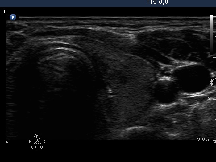
Left lobe, horizontal scan. This lobe has a similar presentation as the right lobe.
.

|
|
|
|
Left lobe, horizontal scan. This lobe has a similar presentation as the right lobe.
.