
Differential diagnostic of thyroid cysts - Case 2. (ultrasonographic picture 7)
|
|

|
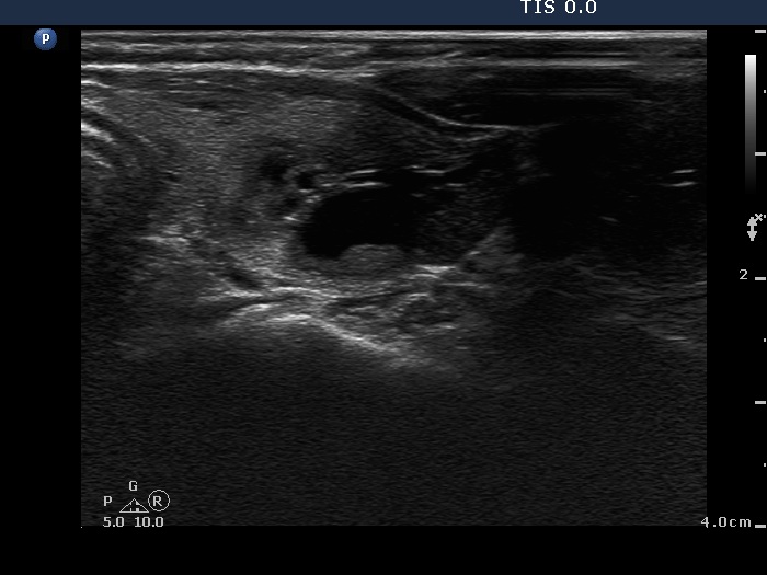
Left lobe, horizontal scan - after aspirating 4.5 ml pale yellow fluid. The hyperechogenic figures correspond to backk wall figures or connective tissue.