
Graves' disease - Case 6.
Follow-up investigation 6 months after first visit (ultrasonographic picture 8)
|
|

|
|
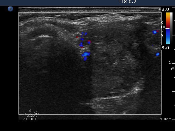
Upper part of the right lobe, horizontal scan, color Doppler mode. The nodule presents a type 1 vascular pattern.