Graves' disease - Case 6. (ultrasonographic picture 5)

|

|

|
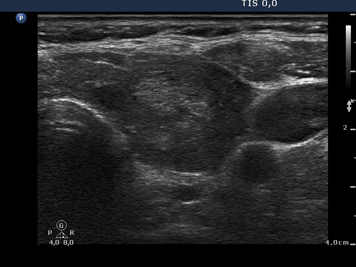
Lower part of the left lobe, horizontal scan. Another hyperechogenic nodule.

|
|
|
|
Lower part of the left lobe, horizontal scan. Another hyperechogenic nodule.


