Graves' disease - Case 6. (ultrasonographic picture 7)
|
|

|
|
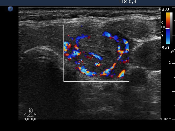
Left lobe, horizontal scan, color Doppler mode. The nodule displays perinodular blood flow.

|
|
|
|
Left lobe, horizontal scan, color Doppler mode. The nodule displays perinodular blood flow.