Graves' disease - Case 30.
21 months after the first visit (ultrasonographic picture 2)
|
|

|
|
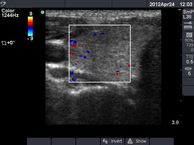
Right lobe, horizontal view, color Doppler mode. The vascularization is normal.

|
|
|
|
Right lobe, horizontal view, color Doppler mode. The vascularization is normal.