Graves' disease - Case 30.
Initial investigation (ultrasonographic picture 3)
|
|

|
|
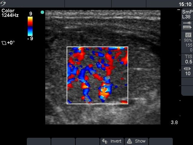
Right lobe, horizontal view, color Doppler mode. The vascularization is increased.

|
|
|
|
Right lobe, horizontal view, color Doppler mode. The vascularization is increased.