
The operated thyroid - Case 19. Recurrent nodules and other lesions in an operated patient
(ultrasonographic picture 6)
|
|

|
|
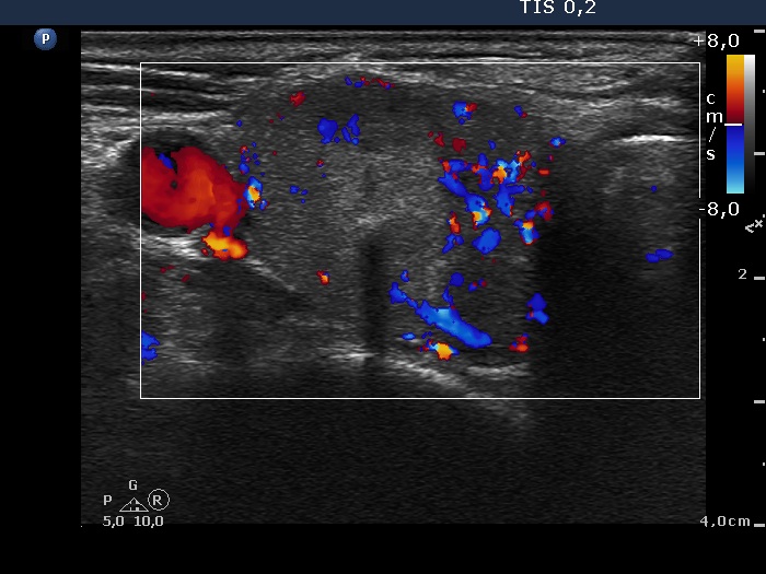
Lower part of the right lobe, horizontal scan, color Doppler mode. The nodule presents type 3 vascular pattern.