
|
|
Medullary carcinoma - Case 4.
|
|


Clinical presentation: a 44-year-old woman was referred for an evaluation of a nodule discovered on screening.
Palpation: no abnormality.
Functional state: euthyroidism with TSH 2.94 mIU/L.
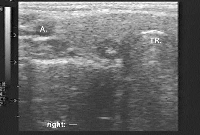
Ultrasonography: demonstrated an 8 mm hypoechogenic nodule with one coarse hyperechogenic granule without dorsal acoustic shadow. ( The patient was evaluated in 1995, therefore the sonographic images are technically out of date. Nevertheless, these are already edifying .)
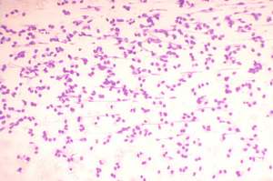
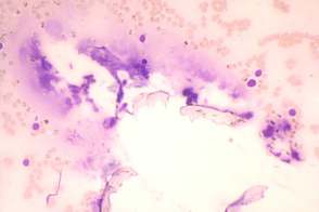
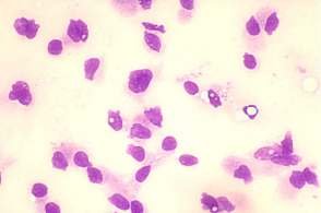
Cytological diagnosis: suspicion of malignancy not otherwise specified.
Immunocytochemistry: LCA, cytokeratin, calcitonin and thyroglobulin reactions were all negative.
Blood test for calcitonin: serum-level of calcitonin was 5.07 pM/L (normal value: 0-3.36).
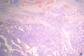
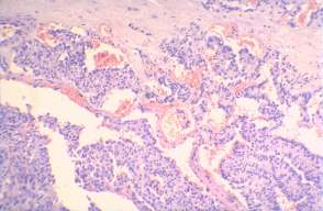
Histopathology: medullary cancer.
Histological specimen courtesy of Dr.Zsolt Orosz, National Institute of Oncology, Budapest, Hungary
Comments:
1. The sonographic pattern is very specific for medullary cancer: cotton-like hyperechogenic patches without acoustic shadow occupying more than 50% of a hypoechogenic nodule.
2. The cytological pattern was not pathognomic. Nevertheless, the lack of normal follicular arrangement is itself suspicious for a tumor not of follicular cell origin.