
Chronic lymphocytic thyroiditis - Case 86. (ultrasonographic picture 7)

|

|

|
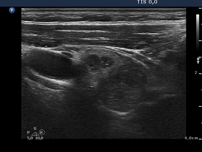
Right side of the isthmus, horizontal scan. The origin of the two small lesions in the ventral part of the image is doubtful, these might be true nodules or only more active foci of thyroiditis. The lateral one presented two bright granules which might be microcalcifications and therefore FNA was performed. The borders of the dorsal and the lateral hypoechogenic areas are ill-defined.


