
The role of complex diagnosis - other examples - Case 4. (ultrasonographic picture 2)
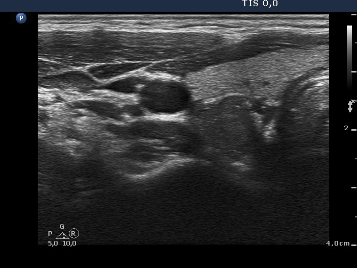 Lower part of the right lobe, horizontal scan. There is another hypoechogenic nodule in the dorsal part of the lobe. It is also suspicious because of the irregular borders. |
