
The role of complex diagnosis - other examples - Case 4. (ultrasonographic picture 6)
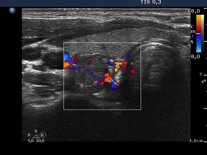 Lower part of the right lobe, horizontal scan, color Doppler mode. This nodule presents type 3 vascular pattern, as well. |
The role of complex diagnosis - other examples - Case 4. (ultrasonographic picture 6)
Lower part of the right lobe, horizontal scan, color Doppler mode. This nodule presents type 3 vascular pattern, as well. |


