
|
|
Ethanol sclerotherapy: thyroid cysts - Case 9
|
|


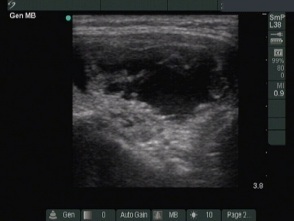


First session (first row of images):
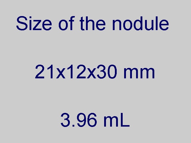
Clinical presentation: a 39-year-old man was referred for a follow-up evaluation of a cystic nodule. We aspirated 1 ml fluid from a nodule with the dimensions of 23x13x28 mm two years ago. The patient noticed an increase in nodule size over the previous months.
Palpation: an elastic nodule in the right lobe.
Functional state: euthyroidism with TSH 0.52 mIU/L.
Ultrasonography: the thyroids were echonormal. There was a dominantly cystic nodule in the right lobe. The lesion displayed signs of perinodular blood flow.
Considering the previous benign cytology and the increase in nodule size we offered ethanol sclerotherapy. 5 mL brown cystic fluid was removed thereafter 2.5 mL ethanol was administered. The treatment caused no complaints.
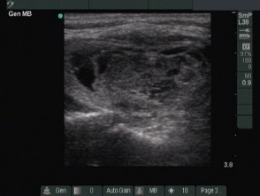
Second session (second row of images):
Clinical presentation: the patient noticed a lump several hours after the therapy which spontaneously withdrew 2 days later.
I aspirated 0.6 mL brown fluid thereafter 2.8 mL ethanol was injected into the nodule. The treatment caused no complaint.
Third session (third row of images):
Clinical presentation: the patient had no complaints after the therapy.
2 mL ethanol was administered into the nodule. The treatment caused a burning, stretching pain which resolved within a minute.
Three months after the last session (fourth row of images):
Clinical presentation: the patient had no complaints. The patient felt a significant shrinkage of the nodule.
Palpation: a small nodule corresponding to the treated lesion.
Functional state: euthyroidism with TSH 1.54 mIU/L.
Ultrasonography. The nodule became much smaller and displayed perinodular blood flow.
4 years after the last session (fifth row of images):
Clinical presentation: the patient had no complaints.
Palpation: a not firm nodule in the lower part of the right lobe.
Functional state: euthyroidism with TSH 1.74 mIU/L.
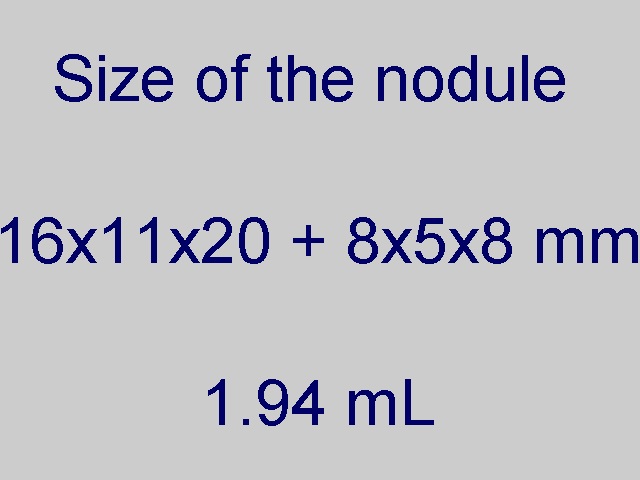
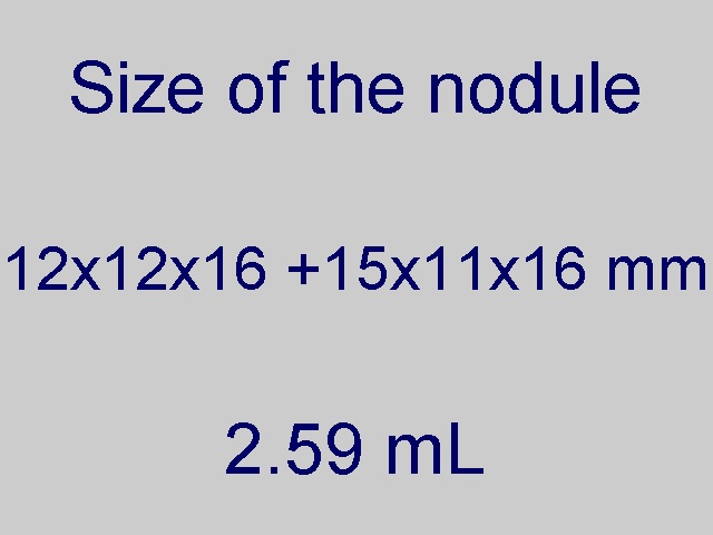
Ultrasonography. Corresponding to the lesion there were three smaller cystic lesions in the right lobe. Although the sum of the volume these areas increased by 25% compared with the previous examination, this figure was less than one-third of the pretreatment size of the lesion.