
Graves' disease - and a concomitant papillary carcinoma - Case 33. (ultrasonographic picture 7)
doi: 10.24390/thyrocase1750.07
|
|

|
|
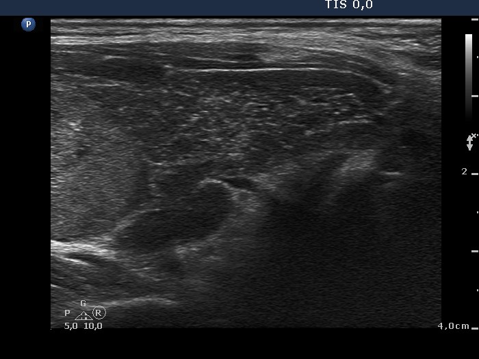
Lower pole of the left lobe, longitudinal scan. In the upper part of the lobe (left in the image) we can see the lower part of the nodule, while in the lower part of the lobe (right in the image) the extranodular parenchyma can be identified.