
Graves' disease - Case 34. (ultrasonographic picture 8)
doi: 10.24390/thyrocase1771.08
|
|

|
|
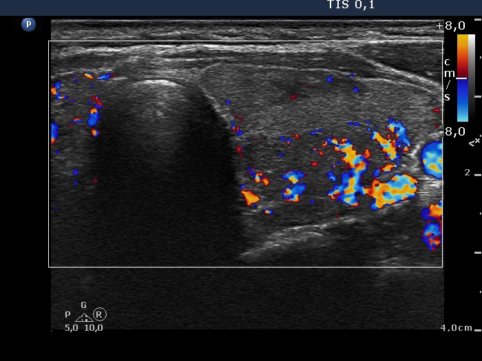
Lower part of the left lobe, horizontal scan, color Doppler mode. The moderately hypoechogenic area presents an irregularly increased vascularization.