
|
|
Medullary carcinoma - Case 8.
|
|


Clinical presentation: A 49-year-old woman was referred for evaluation of a thyroid nodule which was discovered by herself 3 months ago
Palpation. A firm nodule was in the left lobe. Above the left lobe there was an enlarged lymph node.
Functional state: euthyroidism with TSH-level 0.44 mIU/L.
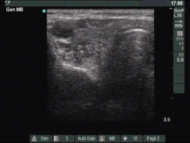
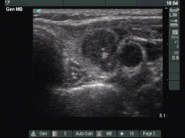
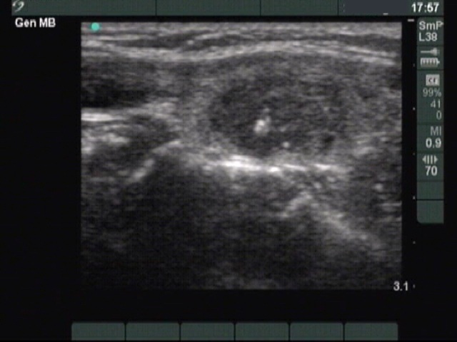
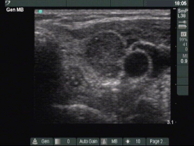
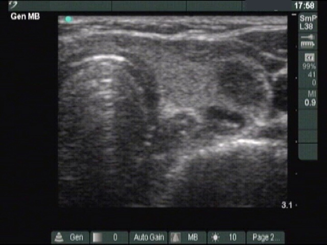
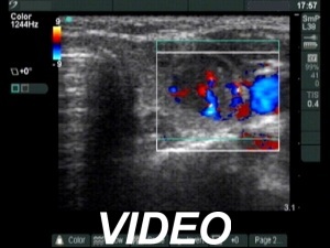
Ultrasonography: There were three hypoechogenic lesions wich has hyperechogenic granules larger than foci of microcalcification. There was a lymph node above the left lobe. The node lacked a regular hilum and was round
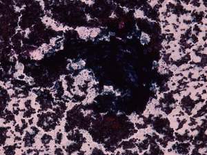
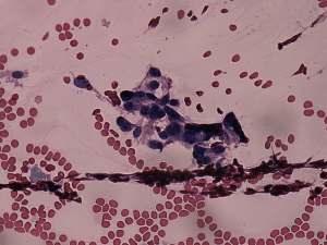
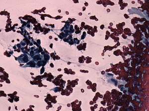
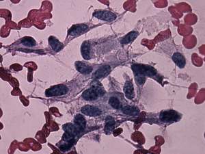
Cytological picture. There was no colloid in the background. There were only a few clusters on the smear within a bloody background. The largest cluster was composed of cells which morphology could not be analyzed. Two other groups were composed of elongated cells. The latter were suspicious because they did not form groups resembling follicles.
The cytological picture itself was not enough for making a diagnosis. But the cytology in combination with the US picture was suspicious for medullary cancer.
Blood test for calcitonin with the result of 2410 pg/mL (normal value: 2-10).
Combined preoperative diagnosis: medullary cancer.
Histopathology revealed multifocal medullary cancer with metastasis to the lymph node of the neck.
Comment. The coarse and large hyperechogenic figures seen on ultrasonography correspond to amyloid deposits. In contrast with coarse calcification, there is no acoustic shadow dorsal to amyloid. The foci of amyloid are much larger than a microcalcification. The specificity of this type of hyperechogenic figures is around 50%.