
Medullary carcinoma - Case 1. (ultrasonographic picture 5)
|
|

|
|
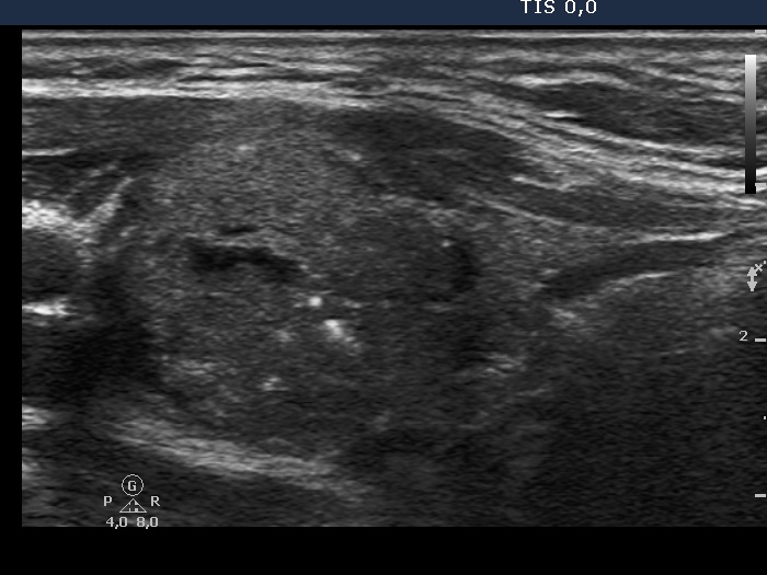
Left lobe, longitudinal scan. Note the various types of hyperechogenic figures within the nodule. The larger bright spots exceed the size of a microcalcification.