
Differentiation of discrete lesions - case 437 (ultrasonographic picture 5)
doi: 10.24390/thyrocase437ln.05
|
|

|
|
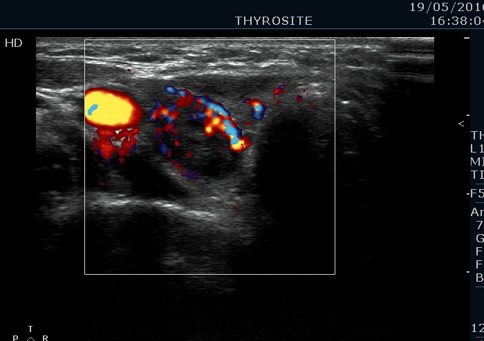
Lower part of the right lobe, horizontal view, color Doppler mode. This is a suspicious vascular pattern.