
The role of complex diagnosis - oxyphilic lesions - Case 7. (ultrasonographic picture 2)
|
|

|
|
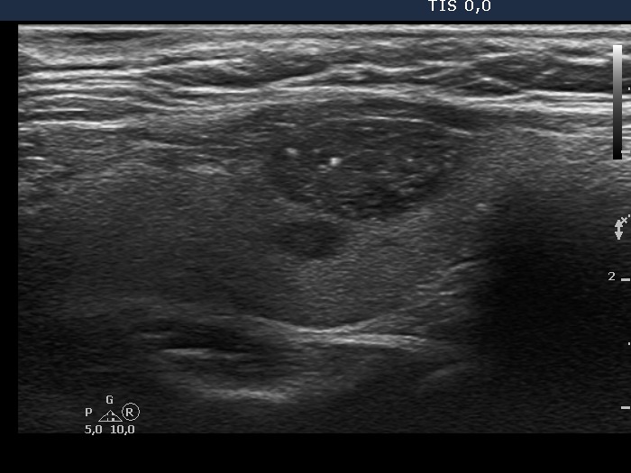
Right lobe, longitudinal scan. The nodule does not display halo sign. There are two larger granules in the lesion. The brighter in the central part could be a microcalcification but as the video proves, similarly bright hyperechogenic lines are also present. So, this large and bright granule is very likely also a presentation of connective tissue.