
|
|
Chronic lymphocytic thyroiditis - Case 7.
|
|


Clinical presentation: a 41-year-old woman was referred for an evaluation of suspected hypothyroidism and a nodular goiter detected on ultrasound.
Palpation: both thyroids were firm, no nodule was palpable.
Functional state: hypothyroidism with TSH 8.71 mIU/L, FT4 8.93 pM/L.
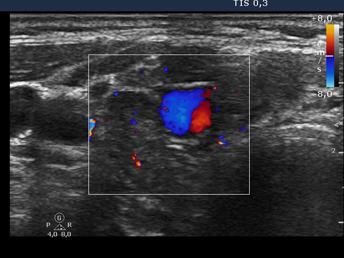
Ultrasonography: the thyroids were moderately hypoechogenic and contained numerous more hypoechogenic areas. These did not fit nodule. There was a relatively large vessel in the central part of the right lobe.
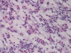
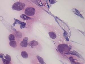
FNAC: was performed from an inhomogeneous hypoechogenic area in the upper part of the right lobe. Cytology disclosed Hashimoto's thyroiditis.