Graves' disease - Case 24. (ultrasonographic picture 4)
|
|

|
|
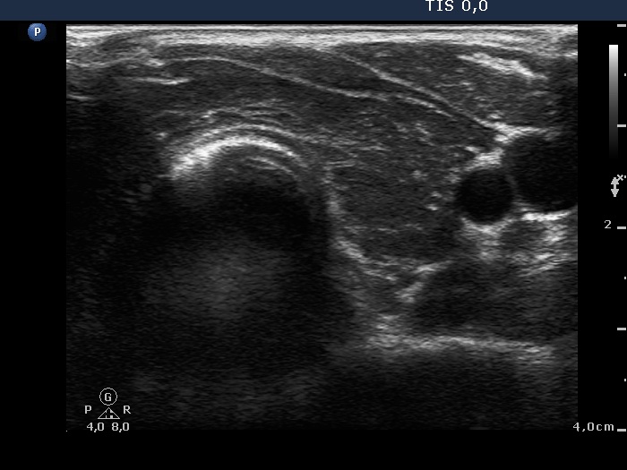
Left lobe, horizontal scan. Note fibrotic changes.

|
|
|
|
Left lobe, horizontal scan. Note fibrotic changes.