
Graves' disease - Case 21. (ultrasonographic picture 5)
|
|

|
|
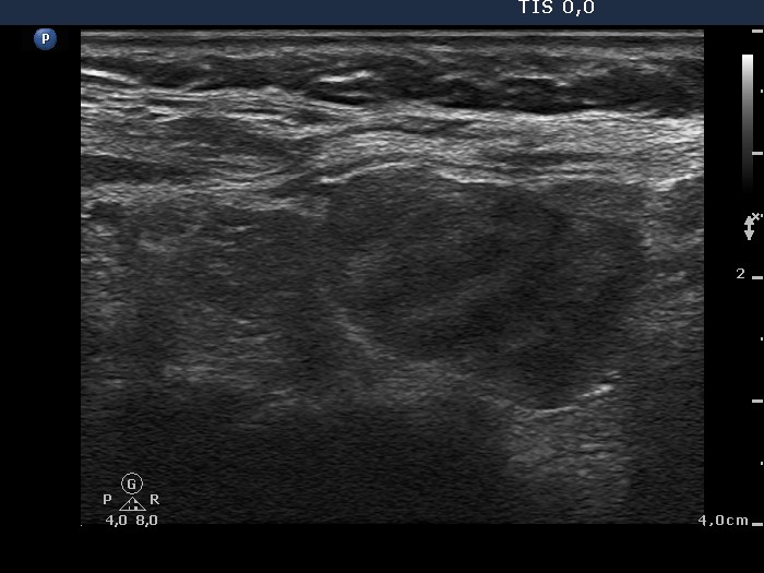
Left lobe, longitudinal scan. The hypoechogenic area is divided to a smaller upper (left in the image) and a larger lower part (right in the image) by connective tissue.