
Differentiation of discrete lesions - case 790 (ultrasonographic picture 6)
doi: 10.24390/thyrocase790.06
|
|

|
|
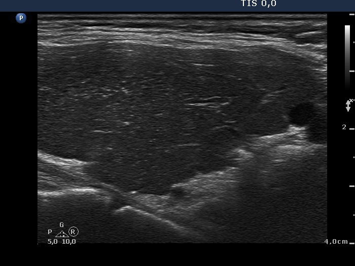
Lower part of the left lobe, longitudinal view. This pattern is very edifying. There are numerous bright echogenic foci which are in this case presentations of connective tissue because of the synchronous presence of echogenic lines. Naturally, in this case the presence of echogenic granules does not cause any differential diagnostic problem. However, a similar presentation of echogenic punctate foci in the event of a nodule is not infrequently misinterpreted as microcalcification.