
|
|
Differential diagnostic of thyroid cysts - Case 10.
|
|


Clinical presentation: A 66-year-old woman was referred for follow-up of a nodule discovered on screening 6 years ago. The woman had no complaints.
Palpation: no abnormality.
Functional state: euthyroidism with TSH 1.72 mIU/L.
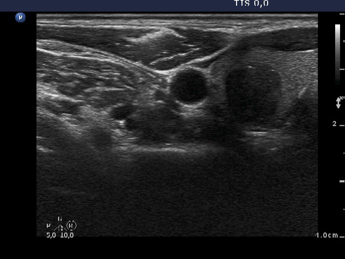
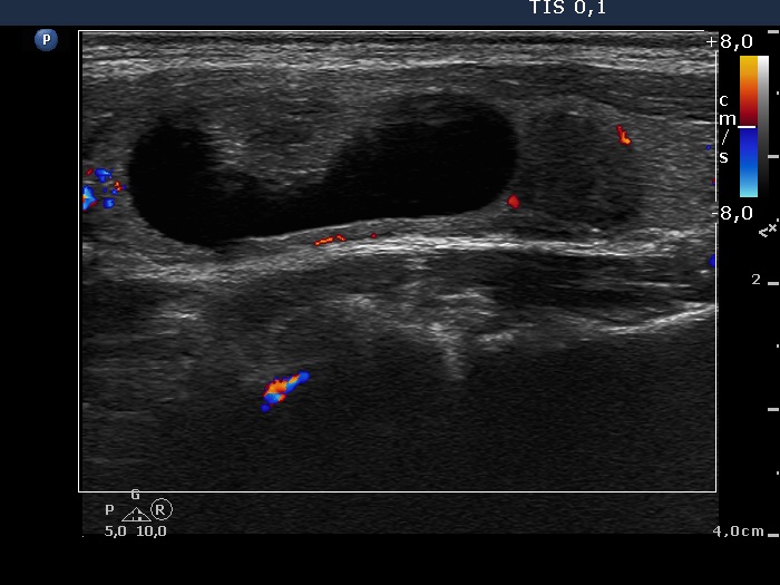
Ultrasonography. The thyroid was echonormal. There was a spongiform-type cyst in the right lobe. The small cystic areas of the lesion exhibited posterior back wall enhancement. The vascularization was scanty. Compared with the former examination, the nodule remained just as large. There was another nodule in the left lobe which was not described on previous ultrasound report. This lesion was hypoechogenic, presented hyperechogenic granules and showed taller-than-wide sign.
Aspiration cytology was performed from the lower, hypoechogenic lesion and resulted in benign colloid goiter.
Suggestion ultrasound in five years.
Comment. Note the typical presentation of posterior back wall enhancement.