Consecutive patients with Hashimoto's thyroiditis - Case 7. (ultrasonographic picture 6)
|
|

|
|
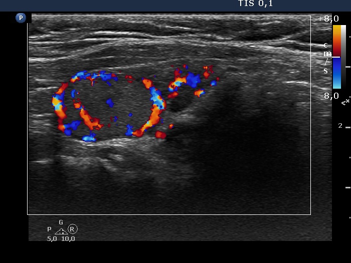
Left lobe, longitudinal scan, color Doppler mode. The lesion has perinodular and intranodular blood flow.

|
|
|
|
Left lobe, longitudinal scan, color Doppler mode. The lesion has perinodular and intranodular blood flow.