
Anaplastic carcinoma - Case 11. (cytologic picture 7)
|
|

|

|
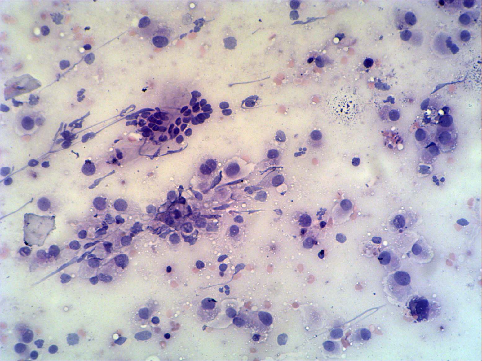
Smear 2. Pap-staining, 200x. Beside enlarged oxyphilic cells, there is a typical cell group in the centre of the image. Note nuclear debris. This pattern is identical with that observed in the event of a Hashimoto thyroiditis.