
|
|
Secondary thyroid carcinomas - Case 2.Metastasis of a urinary bladder carcinoma |
|


Clinical presentation: a 58-year-old man was referred for an evaluation of a newly discovered nodule. Ha was operated on for urinary bladder cancer 6 months earlier. On routine examination suspicious foci in the liver and a thyroid nodule were discovered.
Palpation: a very hard, fixed mass in the right thyroid and enlarged lymph node in the right side of the neck.
Functional state: euthyroidism (TSH-level 1.12 mIU/L).
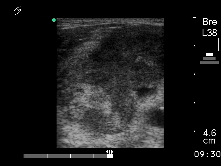
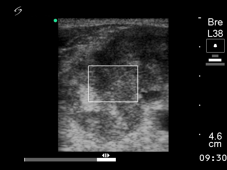
Ultrasonography: the right lobe was enlarged and consisted of multiple hypoechogenic nodules. Multiple enlarged lymph nodes were detected on the right side of the neck.
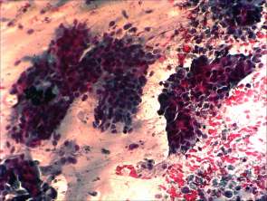
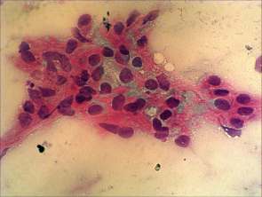
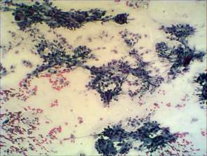
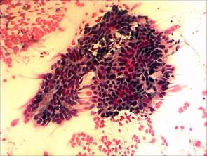
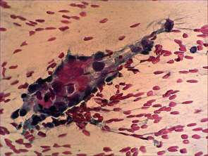
Cytological picture: no colloid in the background. Hyperchromatic epithelial columnar cells were found with clear cytoplasm. They were located in irregular cell groups with loss of polarity and nuclear crowding.
Cytological diagnosis: a pattern corresponding to metastasis of the urinary bladder cancer.