
|
|
The role of complex diagnosis - non-diagnostic cytologies - not diagnostic cytology - Case 4.
|
|


Clinical presentation: a 38-year-old woman was referred for evaluation of a nodule which was discovered by screening.
Palpation: an elastic nodule in the right lobe.
Functional state: euthyroidism with TSH-level 1.38 mIU/L.
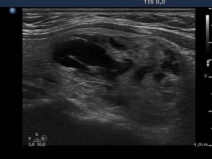
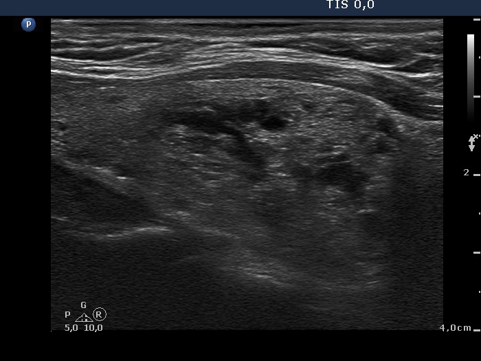
Ultrasonography: the thyroid was echonormal. There were two mixed nodules in the thyroid, one larger in the right and another smaller one in the left lobe. The vascular type of the nodules were not specific, they did not presented a halo.
After aspirating 7 mL brown fluid from the right nodule, the lesion presented a type 2 vascular pattern.
Cytology was performed. Only macrophages and blood cells were on the smear.
Final diagnosis: nodular goiter with not greater than 1% risk of malignancy. Considering the ultrasound presentation we told the patient that the risk of malignancy is not greater than 1% and offered regular follow-up examination.
6 months later the cyst has refilled and the patient decided to undergo surgery.
A right lobectomy was performed. Histopathology: benign cystadenoma in the right thyroid.
Comment. In the event of a relatively large solitary nodule presenting halo and type 2 vascular pattern, the diagnosis of a follicular tumor is very likely. In contrast with an adenoma, a follicular carcinoma only exceptionally undergoes spongiform-type or central-type cystic degeneration.