
The operated thyroid - Case 20. (ultrasonographic picture 5)
A previously operated patient with a newly developed Graves' disease

|

|

|
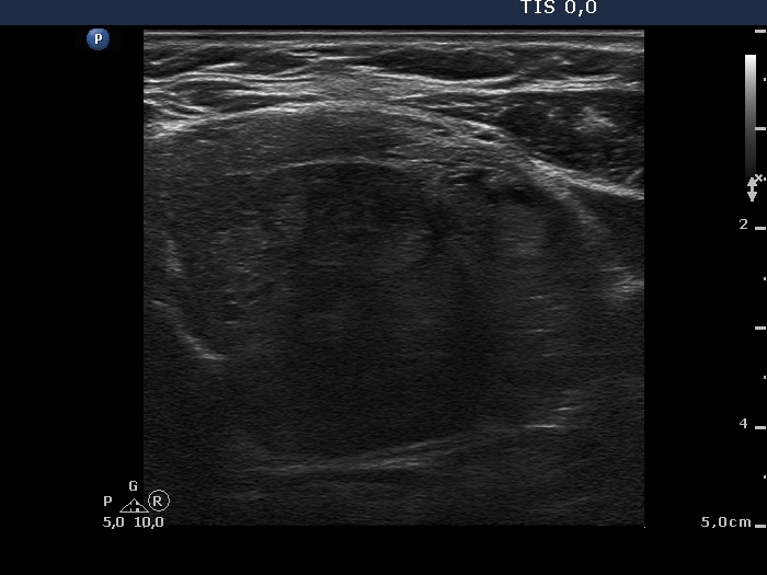
Left lobe, horizontal scan. The pattern is similar to that demonstrated in the right lobe except for the presence of small hyperechogenic lesions of regular geometrical shape.


