
Graves' disease - Case 14. (ultrasonographic picture 4)
|
|

|
|
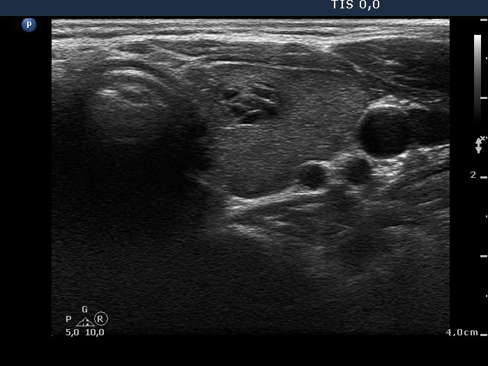
Left lobe, horizontal scan. There is a cystic lesion in the central part of the minimally-moderately hypoechogenic lobe. The bright spots sit on fibrotic bounds - these are not microcalcifications but optical artifacts caused by posterior back wall enhancement.