
|
|
100 consecutive patients with papillary carcinoma - Case 2.
|
|


Clinical data: A 24-year-old woman requested an evaluation of her goiter known for several years. She has been treated with levothyroxine in order to decrease the size of her nodule. Except for ultrasound and a normal TSH no other tests were performed.
Palpation: The right lobe was firm and nodular.
Functional state: euthyroidism with subnormal TSH on daily 75 microgram levothyroxine: TSH 0.13 mIU/L, FT4 1.23 ng/dL.
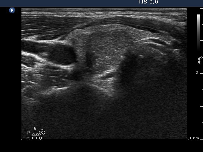
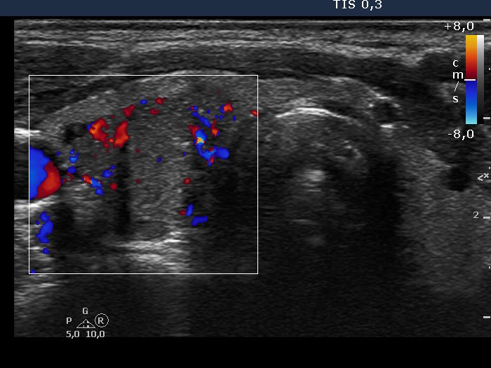
Ultrasonography. The thyroid was echonormal. There was a hypoechogenic nodule presenting irregular lobulated margin and intranodular hyperechogenic figures including microcalcifications. The nodule had an irregularly increased intranodular vascularization. A small, moderately hypoechogenic inhomogeneous lesion was found in the left lobe.
Cytology was performed from both lesions and resulted in papillary carcinoma and benign colloid goiter, right nodule and left nodule, respectively.
Histopathology disclosed papillary carcinoma in the right nodule while hyperplastic nodule in the left nodule.
Comment. It is worth to analyze intranodular hyperechogenic figures both in the images and in the video. The right nodule had not hyperechogenic granules but also similarly bright echogenic lines. Therefore, it is equivocal whether these figures are microcalcifications or presentations of fibrosis.