
100 consecutive patients with papillary carcinoma - Case 3. (ultrasonographic picture 3)
|
|

|
|
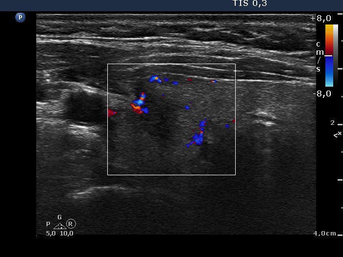
Right lobe, horizontal scan, color Doppler mode. The lesion presents signs of increased intranodular blood flow.