
|
|
100 consecutive patients with papillary carcinoma - Case 5.
|

|


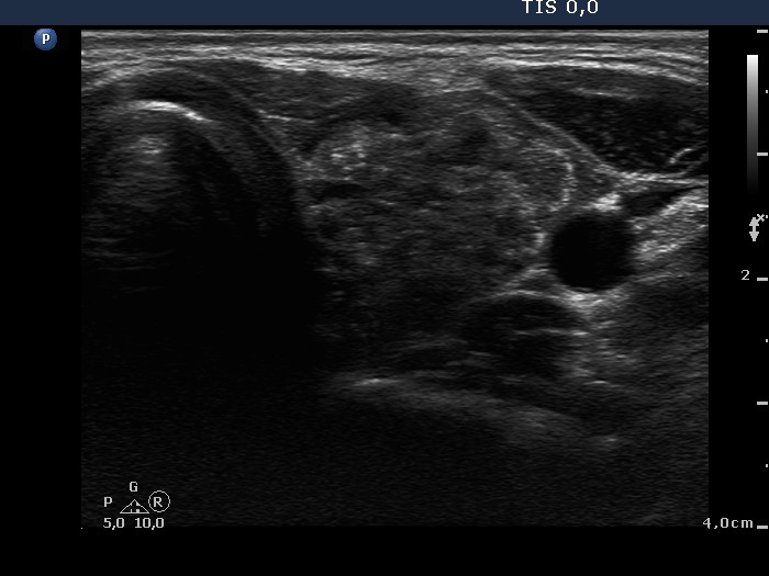
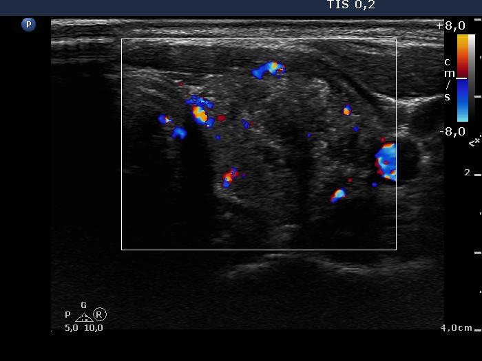
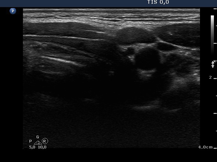



Clinical data: A 17-year-old girl was referred for evaluation of a nodule discovered by herself for 3 weeks.
Palpation: a very firm nodule with uneven surface in the left lobe.
Functional state: euthyroidism (TSH-level 1.89 mIU/L).
Ultrasonography: The thyroid was echonormal and had a small insignificant moderately hypoechogenic lesion in the right lobe and a hypoechogenic nodule in the left lobe. The latter contained numerous microcalcifications and coarse calcification, as well. The intranodular blood flow was a little bit increased. We found an enlarged lymph node in the left side of the neck.
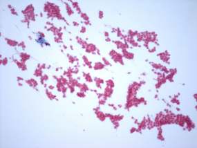
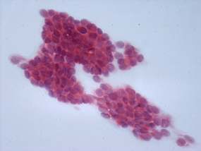
Cytological picture: There was no colloid in the background. Irregular clusters presented with nuclear crowding and overlapping, there were several typical papillary fronds. The thyrocytes were enlarged. I have found only a few grooves and one inclusion in the smear.
Cytological diagnosis: suspicion of papillary carcinoma.
Histopathology disclosed a hyperplastic nodule in the right lobe while a papillary carcinoma destroying the borders of the organ in the left lobe. Metastatic lymph nodes were found in both sides of the neck.


