
Differentiation of discrete lesions - case p016 (ultrasonographic picture 9)
doi: 10.24390/thyrocaseconp016ln.09

|

|

|
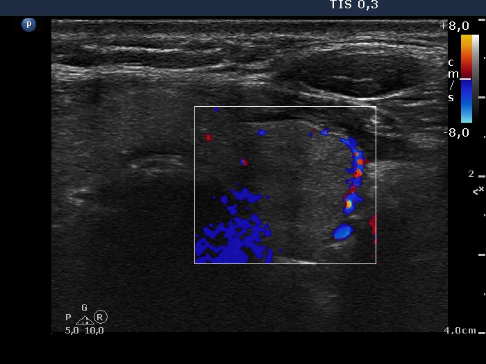
Left lobe, horizontal view, color Doppler mode. There are signs of perinodular blood flow.


