
|
|
100 consecutive patients with papillary carcinoma - Case 20.
|
|


Clinical data: A 43-year-old woman was referred for follow-up examination. ve met the patient for the first time first 13 years ago when a hypoechogenic lesion was found on ultrasound examination. The dimensions of the lesion were 7x7x9 mm, width, depth, length, respectively. A repeat ultrasound was suggested in 5 years.
Palpation: no abnormality.
Functional state: euthyroidism (TSH 0.67 mIU/L, anti-TPO 8 U/mL).
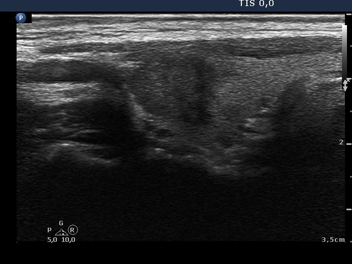
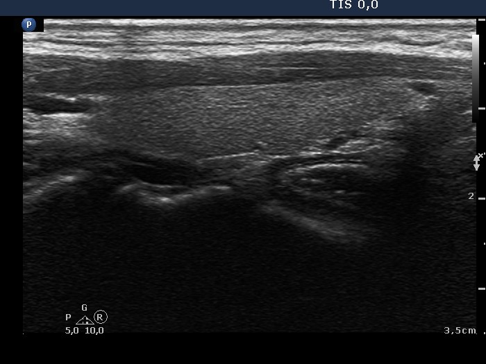
Ultrasonography. The thyroid was echonormal. There was a hypoechogenic lesion in the upper, ventrolateral part of the right lobe. The lesion presented partly lobular, partly blurred surface. The dimensions of the nodule were 9x12x13 mm, width, depth, length, respectively, it means that the volume of the lesion has increased by almost 100% in the past 13 years.
Cytology resulted in papillary carcinoma.
Histopathology disclosed papillary carcinoma in the right lobe.